 Movie
Movie Controller

[English] 日本語
 Yorodumi
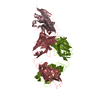
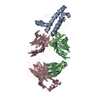
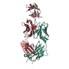
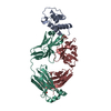
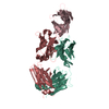
- PDB-5xxy: Crystal structure of PD-L1 complexed with atezolizumab fab at 2.9A -
+ Open data

- Basic information
| Entry | Database: PDB / ID: 5xxy | ||||||
|---|---|---|---|---|---|---|---|
| Title | Crystal structure of PD-L1 complexed with atezolizumab fab at 2.9A | ||||||
 Components Components |
| ||||||
 Keywords Keywords | IMMUNE SYSTEM / PD-L1 / ATEZOLIZUMAB | ||||||
| Function / homology |  Function and homology information Function and homology informationnegative regulation of tumor necrosis factor superfamily cytokine production / positive regulation of activated CD8-positive, alpha-beta T cell apoptotic process / negative regulation of CD8-positive, alpha-beta T cell activation / TRIF-dependent toll-like receptor signaling pathway / negative regulation of T cell mediated immune response to tumor cell / negative regulation of CD4-positive, alpha-beta T cell proliferation / STAT3 nuclear events downstream of ALK signaling / negative regulation of interleukin-10 production / negative regulation of T cell activation / negative regulation of activated T cell proliferation ...negative regulation of tumor necrosis factor superfamily cytokine production / positive regulation of activated CD8-positive, alpha-beta T cell apoptotic process / negative regulation of CD8-positive, alpha-beta T cell activation / TRIF-dependent toll-like receptor signaling pathway / negative regulation of T cell mediated immune response to tumor cell / negative regulation of CD4-positive, alpha-beta T cell proliferation / STAT3 nuclear events downstream of ALK signaling / negative regulation of interleukin-10 production / negative regulation of T cell activation / negative regulation of activated T cell proliferation / negative regulation of type II interferon production / positive regulation of interleukin-10 production / Co-inhibition by PD-1 / negative regulation of T cell receptor signaling pathway / negative regulation of T cell proliferation / T cell costimulation / response to cytokine / positive regulation of T cell proliferation / recycling endosome membrane / actin cytoskeleton / cellular response to lipopolysaccharide / early endosome membrane / adaptive immune response / transcription coactivator activity / cell surface receptor signaling pathway / immune response / receptor ligand activity / external side of plasma membrane / signal transduction / extracellular exosome / nucleoplasm / plasma membrane Similarity search - Function | ||||||
| Biological species |  Homo sapiens (human) Homo sapiens (human) | ||||||
| Method |  X-RAY DIFFRACTION / SYNCHROTRON / MOLECULAR REPLACEMENT / Resolution: 2.9 Å X-RAY DIFFRACTION / SYNCHROTRON / MOLECULAR REPLACEMENT / Resolution: 2.9 Å | ||||||
 Authors Authors | Zhou, A. / Zhang, F. | ||||||
 Citation Citation | Journal: Oncotarget / Year: 2017 Title: Structural basis of the therapeutic anti-PD-L1 antibody atezolizumab. Authors: Zhang, F. / Qi, X. / Wang, X. / Wei, D. / Wu, J. / Feng, L. / Cai, H. / Wang, Y. / Zeng, N. / Xu, T. / Zhou, A. / Zheng, Y. | ||||||
| History |
|
- Structure visualization
| Structure viewer | Molecule: MolmilJmol/JSmol |
|---|
- Downloads & links
-Download
| PDBx/mmCIF format | 5xxy.cif.gz | 292.2 KB | Display | PDBx/mmCIF format |
|---|---|---|---|---|
| PDB format | pdb5xxy.ent.gz | 241.9 KB | Display | PDB format |
| PDBx/mmJSON format | 5xxy.json.gz | Tree view | PDBx/mmJSON format | |
| Others |  Other downloads Other downloads |
-Validation report
| Arichive directory | https://data.pdbj.org/pub/pdb/validation_reports/xx/5xxyftp://data.pdbj.org/pub/pdb/validation_reports/xx/5xxy | HTTPS FTP |
|---|
-Related structure data


-Links
 PDBj
PDBj


- Assembly
| Deposited unit | 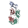
| ||||||||
|---|---|---|---|---|---|---|---|---|---|
| 1 |
| ||||||||
| Unit cell |
|
-Components
| #1: Antibody | Mass: 25677.572 Da / Num. of mol.: 1 Source method: isolated from a genetically manipulated source Source: (gene. exp.) Homo sapiens (human) / Cell line (production host): HEK293S / Production host: Homo sapiens (human) |
|---|---|
| #2: Antibody | Mass: 23386.951 Da / Num. of mol.: 1 Source method: isolated from a genetically manipulated source Source: (gene. exp.) Homo sapiens (human) / Cell line (production host): HEK293S / Production host: Homo sapiens (human) |
| #3: Protein | Mass: 14204.211 Da / Num. of mol.: 1 / Fragment: IgV domain,UNP residues 18-133 Source method: isolated from a genetically manipulated source Source: (gene. exp.) Homo sapiens (human) / Gene: CD274, B7H1, PDCD1L1, PDCD1LG1, PDL1 / Production host:  |
| #4: Water | ChemComp-HOH /  Mass: 18.015 Da / Num. of mol.: 2 / Source method: isolated from a natural source / Formula: H2O Mass: 18.015 Da / Num. of mol.: 2 / Source method: isolated from a natural source / Formula: H2O |
| Has protein modification | Y |
-Experimental details
-Experiment
| Experiment | Method: X-RAY DIFFRACTION / Number of used crystals: 1 |
|---|
- Sample preparation
| Crystal | Density Matthews: 2.26 Å3/Da / Density % sol: 45.53 % |
|---|---|
| Crystal grow | Temperature: 298 K / Method: vapor diffusion, sitting drop / pH: 7 / Details: 2M ammonium sulfate, 0.1M Tris PH 7.0 |
-Data collection
| Diffraction | Mean temperature: 100 K |
|---|---|
| Diffraction source | Source: SYNCHROTRON / Site: SSRF  / Beamline: BL17U1 / Wavelength: 0.9792 Å / Beamline: BL17U1 / Wavelength: 0.9792 Å |
| Detector | Type: MARRESEARCH / Detector: CCD / Date: Apr 30, 2017 |
| Radiation | Protocol: SINGLE WAVELENGTH / Monochromatic (M) / Laue (L): M / Scattering type: x-ray |
| Radiation wavelength | Wavelength: 0.9792 Å / Relative weight: 1 |
| Reflection | Resolution: 2.9→61.72 Å / Num. obs: 11768 / % possible obs: 91 % / Redundancy: 3 % / Net I/σ(I): 4.4 |
| Reflection shell | Resolution: 2.9→3.08 Å / Rmerge(I) obs: 0.573 / Mean I/σ(I) obs: 1.8 / CC1/2: 0.612 / % possible all: 95.3 |
- Processing
| Software |
| ||||||||||||||||||||||||||||||||||||||||
|---|---|---|---|---|---|---|---|---|---|---|---|---|---|---|---|---|---|---|---|---|---|---|---|---|---|---|---|---|---|---|---|---|---|---|---|---|---|---|---|---|---|
| Refinement | Method to determine structure: MOLECULAR REPLACEMENT Starting model: 5JDS,5GGT Resolution: 2.9→61.601 Å / SU ML: 0.33 / Cross valid method: FREE R-VALUE / σ(F): 1.34 / Phase error: 28.96 / Stereochemistry target values: ML
| ||||||||||||||||||||||||||||||||||||||||
| Solvent computation | Shrinkage radii: 0.9 Å / VDW probe radii: 1.11 Å / Solvent model: FLAT BULK SOLVENT MODEL | ||||||||||||||||||||||||||||||||||||||||
| Refinement step | Cycle: LAST / Resolution: 2.9→61.601 Å
| ||||||||||||||||||||||||||||||||||||||||
| Refine LS restraints |
| ||||||||||||||||||||||||||||||||||||||||
| LS refinement shell |
| ||||||||||||||||||||||||||||||||||||||||
| Refinement TLS params. | Method: refined / Origin x: 0.0589 Å / Origin y: -32.9488 Å / Origin z: 17.5613 Å
| ||||||||||||||||||||||||||||||||||||||||
| Refinement TLS group | Selection details: all |
