 Movie
Movie Controller

[English] 日本語
 Yorodumi
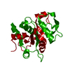
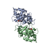
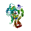
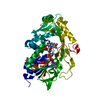
- PDB-1tks: Crystal structure of 3,4-Dihydroxy-2-butanone 4-phosphate Synthas... -
+ Open data

- Basic information
| Entry | Database: PDB / ID: 1tks | ||||||
|---|---|---|---|---|---|---|---|
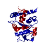
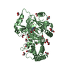
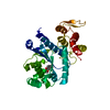
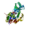
| Title | Crystal structure of 3,4-Dihydroxy-2-butanone 4-phosphate Synthase of Candida albicans | ||||||
 Components Components | 3,4-dihydroxy-2-butanone 4-phosphate synthase | ||||||
 Keywords Keywords | ISOMERASE / Candida albicans / riboflavin biosynthesis / 3 / 4-dihydroxy-2-butanone 4-phosphate synthase / crystal structure synthetic gene | ||||||
| Function / homology |  Function and homology information Function and homology information3,4-dihydroxy-2-butanone-4-phosphate synthase / 3,4-dihydroxy-2-butanone-4-phosphate synthase activity / riboflavin biosynthetic process / mitochondrial intermembrane space / metal ion binding / cytosol Similarity search - Function | ||||||
| Biological species |  Candida albicans (yeast) Candida albicans (yeast) | ||||||
| Method |  X-RAY DIFFRACTION / MOLECULAR REPLACEMENT / Resolution: 1.6 Å X-RAY DIFFRACTION / MOLECULAR REPLACEMENT / Resolution: 1.6 Å | ||||||
 Authors Authors | Echt, S. / Bauer, S. / Steinbacher, S. / Huber, R. / Bacher, A. / Fischer, M. | ||||||
 Citation Citation | Journal: J.Mol.Biol. / Year: 2004 Title: Potential anti-infective targets in pathogenic yeasts: structure and properties of 3,4-dihydroxy-2-butanone 4-phosphate synthase of Candida albicans. Authors: Echt, S. / Bauer, S. / Steinbacher, S. / Huber, R. / Bacher, A. / Fischer, M. | ||||||
| History |
|
- Structure visualization
| Structure viewer | Molecule: MolmilJmol/JSmol |
|---|
- Downloads & links
-Download
| PDBx/mmCIF format | 1tks.cif.gz | 101.5 KB | Display | PDBx/mmCIF format |
|---|---|---|---|---|
| PDB format | pdb1tks.ent.gz | 76.5 KB | Display | PDB format |
| PDBx/mmJSON format | 1tks.json.gz | Tree view | PDBx/mmJSON format | |
| Others |  Other downloads Other downloads |
-Validation report
| Arichive directory | https://data.pdbj.org/pub/pdb/validation_reports/tk/1tksftp://data.pdbj.org/pub/pdb/validation_reports/tk/1tks | HTTPS FTP |
|---|
-Related structure data
| Related structure data |  1tkuC 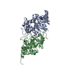 1g57S C: citing same article ( S: Starting model for refinement |
|---|---|
| Similar structure data |

-Links
 PDBj
PDBj- Assembly
| Deposited unit | 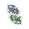
| ||||||||
|---|---|---|---|---|---|---|---|---|---|
| 1 |
| ||||||||
| Unit cell |
|
-Components
| #1: Protein | Mass: 22684.982 Da / Num. of mol.: 2 Source method: isolated from a genetically manipulated source Source: (gene. exp.) Candida albicans (yeast) / Strain: SC5314 / Gene: CARib3 / Plasmid: pNCO-CARib3-syn / Production host:  References: GenBank: 46438026, UniProt: Q5A3V6*PLUS, Isomerases; Intramolecular transferases; Transferring other groups #2: Water | ChemComp-HOH / |  Mass: 18.015 Da / Num. of mol.: 676 / Source method: isolated from a natural source / Formula: H2O Mass: 18.015 Da / Num. of mol.: 676 / Source method: isolated from a natural source / Formula: H2O |
|---|
-Experimental details
-Experiment
| Experiment | Method: X-RAY DIFFRACTION / Number of used crystals: 1 |
|---|
- Sample preparation
| Crystal | Density Matthews: 2.18 Å3/Da / Density % sol: 43 % |
|---|---|
| Crystal grow | Temperature: 293 K / Method: vapor diffusion, sitting drop / pH: 5 Details: Na-Citrat, PEG8000, pH 5.0, VAPOR DIFFUSION, SITTING DROP, temperature 293K |
-Data collection
| Diffraction | Mean temperature: 100 K |
|---|---|
| Diffraction source | Source: ROTATING ANODE / Type: RIGAKU RU200 / Wavelength: 1.542 Å |
| Detector | Type: MARRESEARCH / Detector: IMAGE PLATE / Date: Jul 3, 2003 |
| Radiation | Monochromator: graphite / Protocol: SINGLE WAVELENGTH / Monochromatic (M) / Laue (L): M / Scattering type: x-ray |
| Radiation wavelength | Wavelength: 1.542 Å / Relative weight: 1 |
| Reflection | Resolution: 1.6→20 Å / Num. obs: 43264 / % possible obs: 84.4 % / Redundancy: 3.8 % / Rmerge(I) obs: 0.059 / Net I/σ(I): 27.7 |
| Reflection shell | Resolution: 1.6→1.66 Å / Rmerge(I) obs: 0.221 / % possible all: 81.9 |
- Processing
| Software |
| ||||||||||||
|---|---|---|---|---|---|---|---|---|---|---|---|---|---|
| Refinement | Method to determine structure: MOLECULAR REPLACEMENT Starting model: PDB ENTRY 1G57 Resolution: 1.6→20 Å
| ||||||||||||
| Refinement step | Cycle: LAST / Resolution: 1.6→20 Å
| ||||||||||||
| LS refinement shell | Resolution: 1.6→1.66 Å
|
