 Movie
Movie Controller

[English] 日本語
 Yorodumi
- PDB-1g57: CRYSTAL STRUCTURE OF 3,4-DIHYDROXY-2-BUTANONE 4-PHOSPHATE SYNTHASE -
+ Open data

- Basic information
| Entry | Database: PDB / ID: 1g57 | ||||||
|---|---|---|---|---|---|---|---|
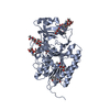
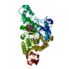
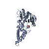
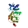
| Title | CRYSTAL STRUCTURE OF 3,4-DIHYDROXY-2-BUTANONE 4-PHOSPHATE SYNTHASE | ||||||
 Components Components | 3,4-DIHYDROXY-2-BUTANONE 4-PHOSPHATE SYNTHASE | ||||||
 Keywords Keywords | ISOMERASE / dihydroxybutanone phosphate synthase / riboflavine biosynthesis / skeletal rearrangement / antimicrobial target / structure-based design | ||||||
| Function / homology |  Function and homology information Function and homology information3,4-dihydroxy-2-butanone-4-phosphate synthase / 3,4-dihydroxy-2-butanone-4-phosphate synthase activity / riboflavin biosynthetic process / manganese ion binding / magnesium ion binding / protein homodimerization activity / identical protein binding / plasma membrane / cytosol Similarity search - Function | ||||||
| Biological species |  | ||||||
| Method |  X-RAY DIFFRACTION / SYNCHROTRON / MAD / Resolution: 1.4 Å X-RAY DIFFRACTION / SYNCHROTRON / MAD / Resolution: 1.4 Å | ||||||
 Authors Authors | Liao, D.-I. / Calabrese, J.C. / Wawrzak, Z. / Viitanen, P.V. / Jordan, D.B. | ||||||
 Citation Citation | Journal: Structure / Year: 2001 Title: Crystal structure of 3,4-dihydroxy-2-butanone 4-phosphate synthase of riboflavin biosynthesis. Authors: Liao, D.I. / Calabrese, J.C. / Wawrzak, Z. / Viitanen, P.V. / Jordan, D.B. | ||||||
| History |
|
- Structure visualization
| Structure viewer | Molecule: MolmilJmol/JSmol |
|---|
- Downloads & links
-Download
| PDBx/mmCIF format | 1g57.cif.gz | 100.5 KB | Display | PDBx/mmCIF format |
|---|---|---|---|---|
| PDB format | pdb1g57.ent.gz | 76.2 KB | Display | PDB format |
| PDBx/mmJSON format | 1g57.json.gz | Tree view | PDBx/mmJSON format | |
| Others |  Other downloads Other downloads |
-Validation report
| Arichive directory | https://data.pdbj.org/pub/pdb/validation_reports/g5/1g57ftp://data.pdbj.org/pub/pdb/validation_reports/g5/1g57 | HTTPS FTP |
|---|
-Related structure data


-Links
 PDBj
PDBj- Assembly
| Deposited unit | 
| ||||||||
|---|---|---|---|---|---|---|---|---|---|
| 1 |
| ||||||||
| Unit cell |
| ||||||||
| Details | The biological assembly is a homodimer. The two molecules in the asymmetric unit represent the biological assembly. The dimer has a 2-fold symmetry. |
-Components
| #1: Protein | Mass: 23335.436 Da / Num. of mol.: 2 Source method: isolated from a genetically manipulated source Source: (gene. exp.) References: UniProt: P0A7J0, Isomerases; Intramolecular transferases; Transferring other groups #2: Chemical | ChemComp-CS /   Mass: 132.905 Da / Num. of mol.: 4 / Source method: obtained synthetically / Formula: Cs Mass: 132.905 Da / Num. of mol.: 4 / Source method: obtained synthetically / Formula: Cs#3: Water | ChemComp-HOH / |  Mass: 18.015 Da / Num. of mol.: 436 / Source method: isolated from a natural source / Formula: H2O Mass: 18.015 Da / Num. of mol.: 436 / Source method: isolated from a natural source / Formula: H2O |
|---|
-Experimental details
-Experiment
| Experiment | Method: X-RAY DIFFRACTION / Number of used crystals: 1 |
|---|
- Sample preparation
| Crystal | Density Matthews: 2.63 Å3/Da / Density % sol: 53.26 % | |||||||||||||||||||||||||
|---|---|---|---|---|---|---|---|---|---|---|---|---|---|---|---|---|---|---|---|---|---|---|---|---|---|---|
| Crystal grow | Temperature: 295 K / Method: vapor diffusion, hanging drop / pH: 7.5 Details: CsCl, Cs(formate), Bis-Tris-propane, pH 7.5, VAPOR DIFFUSION, HANGING DROP, temperature 295K | |||||||||||||||||||||||||
| Crystal grow | *PLUS | |||||||||||||||||||||||||
| Components of the solutions | *PLUS
|
-Data collection
| Diffraction | Mean temperature: 93 K |
|---|---|
| Diffraction source | Source: SYNCHROTRON / Site: APS  / Beamline: 5ID-B / Wavelength: 0.93218 Å / Beamline: 5ID-B / Wavelength: 0.93218 Å |
| Detector | Type: MARRESEARCH / Detector: CCD / Date: Feb 9, 1999 |
| Radiation | Protocol: SINGLE WAVELENGTH / Monochromatic (M) / Laue (L): M / Scattering type: x-ray |
| Radiation wavelength | Wavelength: 0.93218 Å / Relative weight: 1 |
| Reflection | Resolution: 1.4→25 Å / Num. all: 93842 / Num. obs: 1025357 / % possible obs: 100 % / Observed criterion σ(I): -3 / Redundancy: 10.9 % / Rmerge(I) obs: 0.057 / Net I/σ(I): 45.7 |
| Reflection shell | Resolution: 1.4→1.42 Å / Redundancy: 10 % / Rmerge(I) obs: 0.194 / Mean I/σ(I) obs: 10.9 / Num. unique all: 4666 / % possible all: 100 |
| Reflection | *PLUS Num. obs: 93842 / Num. measured all: 1025357 |
| Reflection shell | *PLUS % possible obs: 100 % |
- Processing
| Software |
| ||||||||||||||||||||
|---|---|---|---|---|---|---|---|---|---|---|---|---|---|---|---|---|---|---|---|---|---|
| Refinement | Method to determine structure: MAD / Resolution: 1.4→25 Å / σ(F): 2 / Stereochemistry target values: TNT library
| ||||||||||||||||||||
| Refinement step | Cycle: LAST / Resolution: 1.4→25 Å
| ||||||||||||||||||||
| Refine LS restraints |
|
