 Movie
Movie Controller


+ Open data

- Basic information
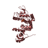
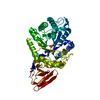
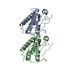
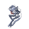
| Entry | Database: PDB / ID: 6lbx | ||||||
|---|---|---|---|---|---|---|---|
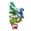
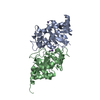
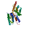
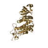
| Title | Crystal structure of HER2 Domain IV and Rb-H2 | ||||||
 Components Components |
| ||||||
 Keywords Keywords | PROTEIN BINDING / kinase | ||||||
| Function / homology |  Function and homology information Function and homology informationnegative regulation of immature T cell proliferation in thymus / ERBB3:ERBB2 complex / ERBB2-ERBB4 signaling pathway / immature T cell proliferation in thymus / GRB7 events in ERBB2 signaling / RNA polymerase I core binding / semaphorin receptor complex / Developmental Lineage of Mammary Stem Cells / ErbB-3 class receptor binding / motor neuron axon guidance ...negative regulation of immature T cell proliferation in thymus / ERBB3:ERBB2 complex / ERBB2-ERBB4 signaling pathway / immature T cell proliferation in thymus / GRB7 events in ERBB2 signaling / RNA polymerase I core binding / semaphorin receptor complex / Developmental Lineage of Mammary Stem Cells / ErbB-3 class receptor binding / motor neuron axon guidance / Sema4D induced cell migration and growth-cone collapse / regulation of microtubule-based process / PLCG1 events in ERBB2 signaling / ERBB2-EGFR signaling pathway / enzyme-linked receptor protein signaling pathway / ERBB2 Activates PTK6 Signaling / ERBB2-ERBB3 signaling pathway / Drug-mediated inhibition of ERBB2 signaling / Resistance of ERBB2 KD mutants to trastuzumab / Resistance of ERBB2 KD mutants to sapitinib / Resistance of ERBB2 KD mutants to tesevatinib / Resistance of ERBB2 KD mutants to neratinib / Resistance of ERBB2 KD mutants to osimertinib / Resistance of ERBB2 KD mutants to afatinib / Resistance of ERBB2 KD mutants to AEE788 / Resistance of ERBB2 KD mutants to lapatinib / Drug resistance in ERBB2 TMD/JMD mutants / neurotransmitter receptor localization to postsynaptic specialization membrane / positive regulation of Rho protein signal transduction / positive regulation of MAP kinase activity / oligodendrocyte differentiation / positive regulation of transcription by RNA polymerase I / ERBB2 Regulates Cell Motility / Developmental Lineage of Mammary Gland Myoepithelial Cells / semaphorin-plexin signaling pathway / neuromuscular junction development / PI3K events in ERBB2 signaling / regulation of angiogenesis / positive regulation of protein targeting to membrane / regulation of ERK1 and ERK2 cascade / Schwann cell development / coreceptor activity / Signaling by ERBB2 / TFAP2 (AP-2) family regulates transcription of growth factors and their receptors / myelination / peptidyl-tyrosine phosphorylation / transmembrane receptor protein tyrosine kinase activity / positive regulation of epithelial cell proliferation / GRB2 events in ERBB2 signaling / positive regulation of cell adhesion / SHC1 events in ERBB2 signaling / cell surface receptor protein tyrosine kinase signaling pathway / cellular response to epidermal growth factor stimulus / basal plasma membrane / Constitutive Signaling by Overexpressed ERBB2 / Downregulation of ERBB2:ERBB3 signaling / positive regulation of translation / phosphatidylinositol 3-kinase/protein kinase B signal transduction / neuromuscular junction / wound healing / Signaling by ERBB2 TMD/JMD mutants / receptor protein-tyrosine kinase / Signaling by ERBB2 ECD mutants / Signaling by ERBB2 KD Mutants / cellular response to growth factor stimulus / receptor tyrosine kinase binding / epidermal growth factor receptor signaling pathway / ruffle membrane / Downregulation of ERBB2 signaling / neuron differentiation / Constitutive Signaling by Aberrant PI3K in Cancer / transmembrane signaling receptor activity / PIP3 activates AKT signaling / myelin sheath / heart development / PI5P, PP2A and IER3 Regulate PI3K/AKT Signaling / positive regulation of cell growth / RAF/MAP kinase cascade / protein tyrosine kinase activity / presynaptic membrane / basolateral plasma membrane / protein phosphorylation / positive regulation of MAPK cascade / early endosome / cell population proliferation / cell surface receptor signaling pathway / signaling receptor complex / apical plasma membrane / endosome membrane / intracellular signal transduction / protein heterodimerization activity / signaling receptor binding / negative regulation of apoptotic process / perinuclear region of cytoplasm / signal transduction / nucleoplasm / ATP binding / membrane / identical protein binding / nucleus Similarity search - Function | ||||||
| Biological species |  Cyclostomata (jawless vertebrates) Cyclostomata (jawless vertebrates) Homo sapiens (human) Homo sapiens (human) | ||||||
| Method |  X-RAY DIFFRACTION / SYNCHROTRON / MOLECULAR REPLACEMENT / Resolution: 2.03 Å X-RAY DIFFRACTION / SYNCHROTRON / MOLECULAR REPLACEMENT / Resolution: 2.03 Å | ||||||
 Authors Authors | Cho, H.S. / Cha, J.S. | ||||||
 Citation Citation | Journal: Comput Struct Biotechnol J / Year: 2021 Title: Computationally-guided design and affinity improvement of a protein binder targeting a specific site on HER2 Authors: Kim, T.Y. / Cha, J.S. / Kim, H. / Choi, Y. / Cho, H.S. / Kim, H.S. | ||||||
| History |
|
- Structure visualization
| Structure viewer | Molecule: MolmilJmol/JSmol |
|---|
- Downloads & links
-Download
| PDBx/mmCIF format | 6lbx.cif.gz | 101 KB | Display | PDBx/mmCIF format |
|---|---|---|---|---|
| PDB format | pdb6lbx.ent.gz | 60.8 KB | Display | PDB format |
| PDBx/mmJSON format | 6lbx.json.gz | Tree view | PDBx/mmJSON format | |
| Others |  Other downloads Other downloads |
-Validation report
| Arichive directory | https://data.pdbj.org/pub/pdb/validation_reports/lb/6lbxftp://data.pdbj.org/pub/pdb/validation_reports/lb/6lbx | HTTPS FTP |
|---|
-Related structure data
| Related structure data | 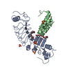 5b4pS S: Starting model for refinement |
|---|---|
| Similar structure data |


-Links
 PDBj
PDBj


- Assembly
| Deposited unit | 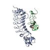
| ||||||||||||
|---|---|---|---|---|---|---|---|---|---|---|---|---|---|
| 1 |
| ||||||||||||
| Unit cell |
|
-Components
| #1: Protein | Mass: 30713.172 Da / Num. of mol.: 1 Source method: isolated from a genetically manipulated source Source: (gene. exp.) Cyclostomata (jawless vertebrates) / Production host:  |
|---|---|
| #2: Protein | Mass: 10845.373 Da / Num. of mol.: 1 / Fragment: Domain IV Source method: isolated from a genetically manipulated source Source: (gene. exp.) Homo sapiens (human) / Gene: ERBB2, HER2, MLN19, NEU, NGL / Production host:   Spodoptera frugiperda (fall armyworm) Spodoptera frugiperda (fall armyworm)References: UniProt: P04626, receptor protein-tyrosine kinase |
| #3: Sugar | ChemComp-NAG /   Type: D-saccharide, beta linking / Mass: 221.208 Da / Num. of mol.: 1 Type: D-saccharide, beta linking / Mass: 221.208 Da / Num. of mol.: 1Source method: isolated from a genetically manipulated source Formula: C8H15NO6 |
| #4: Water | ChemComp-HOH /  Mass: 18.015 Da / Num. of mol.: 148 / Source method: isolated from a natural source / Formula: H2O Mass: 18.015 Da / Num. of mol.: 148 / Source method: isolated from a natural source / Formula: H2O |
| Has ligand of interest | N |
| Has protein modification | Y |
-Experimental details
-Experiment
| Experiment | Method: X-RAY DIFFRACTION / Number of used crystals: 1 |
|---|
- Sample preparation
| Crystal | Density Matthews: 2.33 Å3/Da / Density % sol: 47.25 % |
|---|---|
| Crystal grow | Temperature: 290 K / Method: vapor diffusion / pH: 5.5 Details: 0.1M Sodium Citrate/Citric Acid (pH 5.5), 20% PEG 3000 |
-Data collection
| Diffraction | Mean temperature: 100 K / Serial crystal experiment: N |
|---|---|
| Diffraction source | Source: SYNCHROTRON / Site: Photon Factory  / Beamline: BL-1A / Wavelength: 0.97 Å / Beamline: BL-1A / Wavelength: 0.97 Å |
| Detector | Type: DECTRIS EIGER X 4M / Detector: PIXEL / Date: May 27, 2019 |
| Radiation | Protocol: SINGLE WAVELENGTH / Monochromatic (M) / Laue (L): M / Scattering type: x-ray |
| Radiation wavelength | Wavelength: 0.97 Å / Relative weight: 1 |
| Reflection | Resolution: 2.03→44.89 Å / Num. obs: 25457 / % possible obs: 98.5 % / Redundancy: 2 % / Biso Wilson estimate: 32.17 Å2 / CC1/2: 0.999 / Rmerge(I) obs: 0.0312 / Net I/σ(I): 16.01 |
| Reflection shell | Resolution: 2.03→2.103 Å / Rmerge(I) obs: 0.3777 / Mean I/σ(I) obs: 1.63 / Num. unique obs: 2471 / CC1/2: 0.728 |
- Processing
| Software |
| |||||||||||||||||||||||||||||||||||||||||||||||||||||||||||||||||||||||||||||||||||||||||||||||||||||||||
|---|---|---|---|---|---|---|---|---|---|---|---|---|---|---|---|---|---|---|---|---|---|---|---|---|---|---|---|---|---|---|---|---|---|---|---|---|---|---|---|---|---|---|---|---|---|---|---|---|---|---|---|---|---|---|---|---|---|---|---|---|---|---|---|---|---|---|---|---|---|---|---|---|---|---|---|---|---|---|---|---|---|---|---|---|---|---|---|---|---|---|---|---|---|---|---|---|---|---|---|---|---|---|---|---|---|---|
| Refinement | Method to determine structure: MOLECULAR REPLACEMENT Starting model: 5B4P Resolution: 2.03→44.89 Å / SU ML: 0.2256 / Cross valid method: FREE R-VALUE / σ(F): 1.35 / Phase error: 21.8755
| |||||||||||||||||||||||||||||||||||||||||||||||||||||||||||||||||||||||||||||||||||||||||||||||||||||||||
| Solvent computation | Shrinkage radii: 0.9 Å / VDW probe radii: 1.11 Å | |||||||||||||||||||||||||||||||||||||||||||||||||||||||||||||||||||||||||||||||||||||||||||||||||||||||||
| Displacement parameters | Biso mean: 31.98 Å2 | |||||||||||||||||||||||||||||||||||||||||||||||||||||||||||||||||||||||||||||||||||||||||||||||||||||||||
| Refinement step | Cycle: LAST / Resolution: 2.03→44.89 Å
| |||||||||||||||||||||||||||||||||||||||||||||||||||||||||||||||||||||||||||||||||||||||||||||||||||||||||
| Refine LS restraints |
| |||||||||||||||||||||||||||||||||||||||||||||||||||||||||||||||||||||||||||||||||||||||||||||||||||||||||
| LS refinement shell |
|
