 Movie
Movie Controller

[English] 日本語
 Yorodumi
- PDB-3kva: Structure of KIAA1718 Jumonji domain in complex with alpha-ketogl... -
+ Open data

- Basic information
| Entry | Database: PDB / ID: 3kva | ||||||
|---|---|---|---|---|---|---|---|
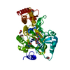
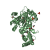
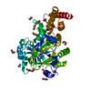
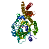
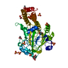
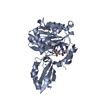
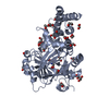
| Title | Structure of KIAA1718 Jumonji domain in complex with alpha-ketoglutarate | ||||||
 Components Components | JmjC domain-containing histone demethylation protein 1D | ||||||
 Keywords Keywords | H3K4ME3 BINDING PROTEIN / TRANSFERASE / Jumonji domain lysine demethylase / Metal-binding / Zinc-finger | ||||||
| Function / homology |  Function and homology information Function and homology informationhistone H3K9me/H3K9me2 demethylase activity / histone H3K27me2/H3K27me3 demethylase activity / [histone H3]-dimethyl-L-lysine9 demethylase / histone H3K36 demethylase activity / histone H4K20 demethylase activity / 2-oxoglutarate-dependent dioxygenase activity / midbrain development / histone H3K9 demethylase activity / histone demethylase activity / transcription coregulator activity ...histone H3K9me/H3K9me2 demethylase activity / histone H3K27me2/H3K27me3 demethylase activity / [histone H3]-dimethyl-L-lysine9 demethylase / histone H3K36 demethylase activity / histone H4K20 demethylase activity / 2-oxoglutarate-dependent dioxygenase activity / midbrain development / histone H3K9 demethylase activity / histone demethylase activity / transcription coregulator activity / HDMs demethylate histones / Signaling by BRAF and RAF1 fusions / chromatin remodeling / iron ion binding / regulation of transcription by RNA polymerase II / nucleolus / positive regulation of DNA-templated transcription / zinc ion binding / nucleoplasm / nucleus Similarity search - Function | ||||||
| Biological species |  Homo sapiens (human) Homo sapiens (human) | ||||||
| Method |  X-RAY DIFFRACTION / SYNCHROTRON / MOLECULAR REPLACEMENT / Resolution: 2.79 Å X-RAY DIFFRACTION / SYNCHROTRON / MOLECULAR REPLACEMENT / Resolution: 2.79 Å | ||||||
 Authors Authors | Horton, J.R. / Upadhyay, A.K. / Qi, H.H. / Zhang, X. / Shi, Y. / Cheng, X. | ||||||
 Citation Citation | Journal: Nat.Struct.Mol.Biol. / Year: 2010 Title: Enzymatic and structural insights for substrate specificity of a family of jumonji histone lysine demethylases. Authors: Horton, J.R. / Upadhyay, A.K. / Qi, H.H. / Zhang, X. / Shi, Y. / Cheng, X. | ||||||
| History |
|
- Structure visualization
| Structure viewer | Molecule: MolmilJmol/JSmol |
|---|
- Downloads & links
-Download
| PDBx/mmCIF format | 3kva.cif.gz | 88.6 KB | Display | PDBx/mmCIF format |
|---|---|---|---|---|
| PDB format | pdb3kva.ent.gz | 65.3 KB | Display | PDB format |
| PDBx/mmJSON format | 3kva.json.gz | Tree view | PDBx/mmJSON format | |
| Others |  Other downloads Other downloads |
-Validation report
| Arichive directory | https://data.pdbj.org/pub/pdb/validation_reports/kv/3kvaftp://data.pdbj.org/pub/pdb/validation_reports/kv/3kva | HTTPS FTP |
|---|
-Related structure data
| Related structure data |  3kv4C  3kv5C  3kv6SC  3kv9C  3kvbC C: citing same article ( S: Starting model for refinement |
|---|---|
| Similar structure data |

-Links
 PDBj
PDBj


- Assembly
| Deposited unit | 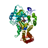
| ||||||||
|---|---|---|---|---|---|---|---|---|---|
| 1 |
| ||||||||
| 2 | 
| ||||||||
| Unit cell |
|
-Components
| #1: Protein | Mass: 46081.043 Da / Num. of mol.: 1 / Fragment: UNP Residues 92-488 Source method: isolated from a genetically manipulated source Details: GST-fusion / Source: (gene. exp.) Homo sapiens (human) / Gene: JHDM1D, KIAA1718 / Production host:  | ||||||
|---|---|---|---|---|---|---|---|
| #2: Chemical |   Mass: 55.845 Da / Num. of mol.: 2 / Source method: obtained synthetically / Formula: Fe Mass: 55.845 Da / Num. of mol.: 2 / Source method: obtained synthetically / Formula: Fe#3: Chemical | ChemComp-OXY / |   Mass: 31.999 Da / Num. of mol.: 1 / Source method: obtained synthetically / Formula: O2 Mass: 31.999 Da / Num. of mol.: 1 / Source method: obtained synthetically / Formula: O2#4: Chemical | ChemComp-AKG / |   Mass: 146.098 Da / Num. of mol.: 1 / Source method: obtained synthetically / Formula: C5H6O5 Mass: 146.098 Da / Num. of mol.: 1 / Source method: obtained synthetically / Formula: C5H6O5#5: Water | ChemComp-HOH / |  Mass: 18.015 Da / Num. of mol.: 50 / Source method: isolated from a natural source / Formula: H2O Mass: 18.015 Da / Num. of mol.: 50 / Source method: isolated from a natural source / Formula: H2O |
-Experimental details
-Experiment
| Experiment | Method: X-RAY DIFFRACTION / Number of used crystals: 1 |
|---|
- Sample preparation
| Crystal | Density Matthews: 2.78 Å3/Da / Density % sol: 55.72 % Description: The Structure Factor File contains Friedel pairs |
|---|---|
| Crystal grow | Temperature: 289 K / Method: vapor diffusion, hanging drop / pH: 6.4 Details: 17-20% (v/v) polyethylene glycol 5000 MME, 0.2 M CaCl2, and 0.1 M BisTris pH 6.4, VAPOR DIFFUSION, HANGING DROP, temperature 289K |
-Data collection
| Diffraction | Mean temperature: 100 K |
|---|---|
| Diffraction source | Source: SYNCHROTRON / Site: APS  / Beamline: 22-ID / Wavelength: 1.27046 Å / Beamline: 22-ID / Wavelength: 1.27046 Å |
| Detector | Type: MARMOSAIC 300 mm CCD / Detector: CCD / Date: Apr 20, 2008 |
| Radiation | Protocol: SINGLE WAVELENGTH / Monochromatic (M) / Laue (L): M / Scattering type: x-ray |
| Radiation wavelength | Wavelength: 1.27046 Å / Relative weight: 1 |
| Reflection | Resolution: 2.79→34.4 Å / Num. obs: 26944 / % possible obs: 99.5 % / Observed criterion σ(F): 0 / Observed criterion σ(I): 0 / Redundancy: 7 % / Biso Wilson estimate: 41.5 Å2 / Rmerge(I) obs: 0.088 / Net I/σ(I): 15.5 |
| Reflection shell | Resolution: 2.79→2.89 Å / Redundancy: 7.3 % / Rmerge(I) obs: 0.381 / Mean I/σ(I) obs: 4.1 / Num. unique all: 1360 / % possible all: 99.3 |
- Processing
| Software |
| ||||||||||||||||||||||||||||||||||||
|---|---|---|---|---|---|---|---|---|---|---|---|---|---|---|---|---|---|---|---|---|---|---|---|---|---|---|---|---|---|---|---|---|---|---|---|---|---|
| Refinement | Method to determine structure: MOLECULAR REPLACEMENT Starting model: 3KV6 Resolution: 2.79→34.4 Å / Rfactor Rfree error: 0.008 / Data cutoff high absF: 555080.86 / Data cutoff low absF: 0 / Isotropic thermal model: RESTRAINED / Cross valid method: THROUGHOUT / σ(F): 0 / Stereochemistry target values: Engh & Huber
| ||||||||||||||||||||||||||||||||||||
| Solvent computation | Solvent model: FLAT MODEL / Bsol: 36.4081 Å2 / ksol: 0.35 e/Å3 | ||||||||||||||||||||||||||||||||||||
| Displacement parameters | Biso mean: 47.8 Å2
| ||||||||||||||||||||||||||||||||||||
| Refine analyze |
| ||||||||||||||||||||||||||||||||||||
| Refinement step | Cycle: LAST / Resolution: 2.79→34.4 Å
| ||||||||||||||||||||||||||||||||||||
| Refine LS restraints |
| ||||||||||||||||||||||||||||||||||||
| LS refinement shell | Resolution: 2.79→2.89 Å / Rfactor Rfree error: 0.036 / Total num. of bins used: 10
| ||||||||||||||||||||||||||||||||||||
| Xplor file |
|
