 Movie
Movie Controller


+ Open data

- Basic information
| Entry | Database: PDB / ID: 3ptr | ||||||
|---|---|---|---|---|---|---|---|
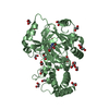
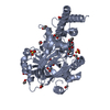
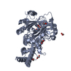
| Title | PHF2 Jumonji domain | ||||||
 Components Components | PHD finger protein 2 | ||||||
 Keywords Keywords | PROTEIN BINDING / alpha-ketoglutarate-Fe2+ dependent dioxygenases / Histone tail BINDING PROTEIN | ||||||
| Function / homology |  Function and homology information Function and homology informationprotein demethylation / negative regulation of rDNA heterochromatin formation / histone H4K20 demethylase activity / Oxidoreductases; Acting on paired donors, with incorporation or reduction of molecular oxygen; With 2-oxoglutarate as one donor, and incorporation of one atom of oxygen into each donor / histone H3K4me3 reader activity / histone H3K9 demethylase activity / histone demethylase activity / transcription initiation-coupled chromatin remodeling / transcription coregulator activity / liver development ...protein demethylation / negative regulation of rDNA heterochromatin formation / histone H4K20 demethylase activity / Oxidoreductases; Acting on paired donors, with incorporation or reduction of molecular oxygen; With 2-oxoglutarate as one donor, and incorporation of one atom of oxygen into each donor / histone H3K4me3 reader activity / histone H3K9 demethylase activity / histone demethylase activity / transcription initiation-coupled chromatin remodeling / transcription coregulator activity / liver development / HDMs demethylate histones / kinetochore / transcription coactivator activity / chromatin remodeling / iron ion binding / regulation of transcription by RNA polymerase II / nucleolus / zinc ion binding / nucleoplasm / nucleus Similarity search - Function | ||||||
| Biological species |  Homo sapiens (human) Homo sapiens (human) | ||||||
| Method |  X-RAY DIFFRACTION / SYNCHROTRON / MOLECULAR REPLACEMENT / Resolution: 1.954 Å X-RAY DIFFRACTION / SYNCHROTRON / MOLECULAR REPLACEMENT / Resolution: 1.954 Å | ||||||
 Authors Authors | Horton, J.R. / Upadhyay, A.K. / Hashimoto, H. / Zhang, X. / Cheng, X. | ||||||
 Citation Citation | Journal: J.Mol.Biol. / Year: 2011 Title: Structural basis for human PHF2 Jumonji domain interaction with metal ions. Authors: Horton, J.R. / Upadhyay, A.K. / Hashimoto, H. / Zhang, X. / Cheng, X. | ||||||
| History |
|
- Structure visualization
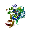
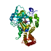
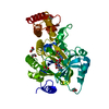
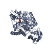
| Structure viewer | Molecule: MolmilJmol/JSmol |
|---|
- Downloads & links
-Download
| PDBx/mmCIF format | 3ptr.cif.gz | 94.8 KB | Display | PDBx/mmCIF format |
|---|---|---|---|---|
| PDB format | pdb3ptr.ent.gz | 70.1 KB | Display | PDB format |
| PDBx/mmJSON format | 3ptr.json.gz | Tree view | PDBx/mmJSON format | |
| Others |  Other downloads Other downloads |
-Validation report
| Arichive directory | https://data.pdbj.org/pub/pdb/validation_reports/pt/3ptrftp://data.pdbj.org/pub/pdb/validation_reports/pt/3ptr | HTTPS FTP |
|---|
-Related structure data
| Related structure data | 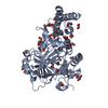 3pu3C 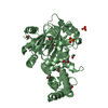 3pu8C  3puaC  3pusC 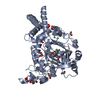 3kv4S C: citing same article ( S: Starting model for refinement |
|---|---|
| Similar structure data |


-Links
 PDBj
PDBj
- Assembly
| Deposited unit | 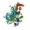
| ||||||||
|---|---|---|---|---|---|---|---|---|---|
| 1 |
| ||||||||
| Unit cell |
|
-Components
| #1: Protein | Mass: 44715.215 Da / Num. of mol.: 1 / Fragment: Jumonji domain (unp residues 60-451) Source method: isolated from a genetically manipulated source Source: (gene. exp.) Homo sapiens (human) / Gene: PHF2, KIAA0662 / Plasmid: pXC870 / Production host:  | ||
|---|---|---|---|
| #2: Chemical | ChemComp-EDO /   Mass: 62.068 Da / Num. of mol.: 19 / Source method: obtained synthetically / Formula: C2H6O2 Mass: 62.068 Da / Num. of mol.: 19 / Source method: obtained synthetically / Formula: C2H6O2#3: Water | ChemComp-HOH / |  Mass: 18.015 Da / Num. of mol.: 247 / Source method: isolated from a natural source / Formula: H2O Mass: 18.015 Da / Num. of mol.: 247 / Source method: isolated from a natural source / Formula: H2O |
-Experimental details
-Experiment
| Experiment | Method: X-RAY DIFFRACTION / Number of used crystals: 1 |
|---|
- Sample preparation
| Crystal | Density Matthews: 2.56 Å3/Da / Density % sol: 51.88 % |
|---|---|
| Crystal grow | Method: vapor diffusion / pH: 6 Details: 18-30% polyethylene glycol 3350 and 100 mM NaCitrate, pH 5.2-6.0, VAPOR DIFFUSION |
-Data collection
| Diffraction | Mean temperature: 100 K |
|---|---|
| Diffraction source | Source: SYNCHROTRON / Site: APS  / Beamline: 22-ID / Wavelength: 1 Å / Beamline: 22-ID / Wavelength: 1 Å |
| Detector | Type: MARMOSAIC 300 mm CCD / Detector: CCD / Date: Apr 26, 2010 |
| Radiation | Protocol: SINGLE WAVELENGTH / Monochromatic (M) / Laue (L): M / Scattering type: x-ray |
| Radiation wavelength | Wavelength: 1 Å / Relative weight: 1 |
| Reflection | Resolution: 1.95→34.85 Å / Num. obs: 33743 / % possible obs: 99.3 % / Observed criterion σ(I): -3 / Redundancy: 6.1 % / Biso Wilson estimate: 22.55 Å2 / Rmerge(I) obs: 0.094 / Net I/σ(I): 18.8 |
| Reflection shell | Resolution: 1.95→2.02 Å / Redundancy: 5.1 % / Rmerge(I) obs: 0.539 / Mean I/σ(I) obs: 3 / Num. unique all: 3254 / % possible all: 97.3 |
- Processing
| Software |
| |||||||||||||||||||||||||||||||||||||||||||||||||||||||||||||||||||||||||||||
|---|---|---|---|---|---|---|---|---|---|---|---|---|---|---|---|---|---|---|---|---|---|---|---|---|---|---|---|---|---|---|---|---|---|---|---|---|---|---|---|---|---|---|---|---|---|---|---|---|---|---|---|---|---|---|---|---|---|---|---|---|---|---|---|---|---|---|---|---|---|---|---|---|---|---|---|---|---|---|
| Refinement | Method to determine structure: MOLECULAR REPLACEMENT Starting model: pdb entry 3KV4 Resolution: 1.954→34.85 Å / SU ML: 0.26 / σ(F): 0.03 / Phase error: 22.38 / Stereochemistry target values: ML
| |||||||||||||||||||||||||||||||||||||||||||||||||||||||||||||||||||||||||||||
| Solvent computation | Shrinkage radii: 0.9 Å / VDW probe radii: 1.11 Å / Solvent model: FLAT BULK SOLVENT MODEL / Bsol: 46.083 Å2 / ksol: 0.345 e/Å3 | |||||||||||||||||||||||||||||||||||||||||||||||||||||||||||||||||||||||||||||
| Displacement parameters |
| |||||||||||||||||||||||||||||||||||||||||||||||||||||||||||||||||||||||||||||
| Refinement step | Cycle: LAST / Resolution: 1.954→34.85 Å
| |||||||||||||||||||||||||||||||||||||||||||||||||||||||||||||||||||||||||||||
| Refine LS restraints |
| |||||||||||||||||||||||||||||||||||||||||||||||||||||||||||||||||||||||||||||
| LS refinement shell |
|
