- PDB-2wwu: Crystal structure of the catalytic domain of PHD finger protein 8 -
+
Open data
ID or keywords:
Loading...
-
Basic information
Entry
Database: PDB / ID: 2wwu
Title
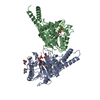
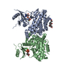
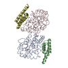
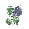
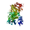
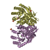
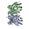
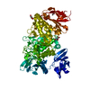
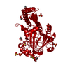
Crystal structure of the catalytic domain of PHD finger protein 8
Components
PHD FINGER PROTEIN 8
Keywords
METAL BINDING PROTEIN / JMJC DOMAIN / EPIGENETICS / METAL-BINDING PROTEIN / HISTONE DEMETHYLASE
Function / homology
Function and homology information
histone H3K36me/H3K36me2 demethylase activity / histone H3K9me/H3K9me2 demethylase activity / histone H3K27me2/H3K27me3 demethylase activity / negative regulation of rDNA heterochromatin formation / [histone H3]-dimethyl-L-lysine9 demethylase / histone H3K36 demethylase activity / histone H4K20 demethylase activity / Oxidoreductases; Acting on paired donors, with incorporation or reduction of molecular oxygen; With 2-oxoglutarate as one donor, and incorporation of one atom of oxygen into each donor / histone H3K4me3 reader activity / histone H3K9 demethylase activity ...histone H3K36me/H3K36me2 demethylase activity / histone H3K9me/H3K9me2 demethylase activity / histone H3K27me2/H3K27me3 demethylase activity / negative regulation of rDNA heterochromatin formation / [histone H3]-dimethyl-L-lysine9 demethylase / histone H3K36 demethylase activity / histone H4K20 demethylase activity / Oxidoreductases; Acting on paired donors, with incorporation or reduction of molecular oxygen; With 2-oxoglutarate as one donor, and incorporation of one atom of oxygen into each donor / histone H3K4me3 reader activity / histone H3K9 demethylase activity / positive regulation of transcription by RNA polymerase I / histone demethylase activity / Condensation of Prophase Chromosomes / transcription coregulator activity / G1/S transition of mitotic cell cycle / brain development / HDMs demethylate histones / nuclear membrane / chromatin remodeling / iron ion binding / chromatin binding / regulation of transcription by RNA polymerase II / nucleolus / positive regulation of DNA-templated transcription / positive regulation of transcription by RNA polymerase II / zinc ion binding / nucleoplasm / nucleus Similarity search - Function
Methane Monooxygenase Hydroxylase; Chain G, domain 1 - #1360 / Jumonji, helical domain / Jumonji helical domain / : / Cupin / JmjC domain, hydroxylase / A domain family that is part of the cupin metalloenzyme superfamily. / JmjC domain / JmjC domain profile. / Zinc finger, PHD-type, conserved site ...Methane Monooxygenase Hydroxylase; Chain G, domain 1 - #1360 / Jumonji, helical domain / Jumonji helical domain / : / Cupin / JmjC domain, hydroxylase / A domain family that is part of the cupin metalloenzyme superfamily. / JmjC domain / JmjC domain profile. / Zinc finger, PHD-type, conserved site / PHD-finger / Zinc finger PHD-type signature. / Methane Monooxygenase Hydroxylase; Chain G, domain 1 / Zinc finger PHD-type profile. / Zinc finger, PHD-finger / Zinc finger, PHD-type / PHD zinc finger / Zinc finger, FYVE/PHD-type / Jelly Rolls / Up-down Bundle / Sandwich / Mainly Beta / Mainly Alpha Similarity search - Domain/homology
ACETATE ION / beta-D-glucopyranose / NICKEL (II) ION / Histone lysine demethylase PHF8 Similarity search - Component
SHEET THE SHEET STRUCTURE OF THIS MOLECULE IS BIFURCATED. IN ORDER TO REPRESENT THIS FEATURE IN ... SHEET THE SHEET STRUCTURE OF THIS MOLECULE IS BIFURCATED. IN ORDER TO REPRESENT THIS FEATURE IN THE SHEET RECORDS BELOW, TWO SHEETS ARE DEFINED.
Resolution: 2.15→33.76 Å / SU ML: 0.34 / σ(F): 1.37 / Phase error: 21.33 / Stereochemistry target values: ML Details: THERE IS DIFFERENCE FO-FC DENSITY AT THE ACTIVE SITE. THE IDENTITY OF THE BOUND LIGAND IS NOT KNOWN, AND HENCE NOT MODELLED.
Rfactor
Num. reflection
% reflection
Rfree
0.2115
1582
5.1 %
Rwork
0.1754
-
-
obs
0.1773
31106
99.76 %
Solvent computation
Shrinkage radii: 0.9 Å / VDW probe radii: 1.11 Å / Solvent model: FLAT BULK SOLVENT MODEL / Bsol: 48.96 Å2 / ksol: 0.361 e/Å3
Displacement parameters
Baniso -1
Baniso -2
Baniso -3
1-
0 Å2
0 Å2
0 Å2
2-
-
0 Å2
0 Å2
3-
-
-
0 Å2
Refinement step
Cycle: LAST / Resolution: 2.15→33.76 Å
Protein
Nucleic acid
Ligand
Solvent
Total
Num. atoms
2900
0
80
168
3148
Refine LS restraints
Refine-ID
Type
Dev ideal
Number
X-RAY DIFFRACTION
f_bond_d
0.008
3057
X-RAY DIFFRACTION
f_angle_d
1.029
4143
X-RAY DIFFRACTION
f_dihedral_angle_d
16.64
1080
X-RAY DIFFRACTION
f_chiral_restr
0.066
465
X-RAY DIFFRACTION
f_plane_restr
0.004
525
LS refinement shell
Resolution (Å)
Rfactor Rfree
Num. reflection Rfree
Rfactor Rwork
Num. reflection Rwork
Refine-ID
% reflection obs (%)
2.1503-2.2197
0.328
156
0.2691
2631
X-RAY DIFFRACTION
100
2.2197-2.299
0.2869
146
0.2399
2668
X-RAY DIFFRACTION
100
2.299-2.391
0.2474
143
0.2107
2646
X-RAY DIFFRACTION
100
2.391-2.4998
0.2593
138
0.1982
2708
X-RAY DIFFRACTION
100
2.4998-2.6316
0.2409
149
0.1927
2641
X-RAY DIFFRACTION
100
2.6316-2.7964
0.2751
144
0.1871
2688
X-RAY DIFFRACTION
100
2.7964-3.0121
0.2123
134
0.195
2692
X-RAY DIFFRACTION
100
3.0121-3.315
0.2205
139
0.1802
2672
X-RAY DIFFRACTION
100
3.315-3.7942
0.2213
148
0.1602
2720
X-RAY DIFFRACTION
100
3.7942-4.7781
0.1624
149
0.1308
2701
X-RAY DIFFRACTION
100
4.7781-33.7643
0.1527
136
0.1554
2757
X-RAY DIFFRACTION
98
Refinement TLS params.
Method: refined / Refine-ID: X-RAY DIFFRACTION
ID
L11 (°2)
L12 (°2)
L13 (°2)
L22 (°2)
L23 (°2)
L33 (°2)
S11 (Å °)
S12 (Å °)
S13 (Å °)
S21 (Å °)
S22 (Å °)
S23 (Å °)
S31 (Å °)
S32 (Å °)
S33 (Å °)
T11 (Å2)
T12 (Å2)
T13 (Å2)
T22 (Å2)
T23 (Å2)
T33 (Å2)
Origin x (Å)
Origin y (Å)
Origin z (Å)
1
1.4555
0.2417
0.8728
0.4391
-0.1071
1.3186
-0.0921
0.2359
0.3245
-0.2049
-0.1259
0.0372
-0.023
0.2467
0.2063
0.3361
0.024
-0.1276
0.3773
-0.0234
0.4165
55.9627
45.9359
10.7862
2
0.5633
-0.3441
0.3365
0.4123
-0.0113
0.3786
-0.274
-0.2274
0.4899
0.0194
-0.0365
0.4017
-0.109
-0.2589
0.1925
0.3909
0.0138
-0.2953
0.4383
-0.0262
0.6234
43.0771
51.7621
9.4966
3
2.4023
0.0659
-0.2279
0.5543
0.0359
1.335
-0.04
-0.1675
0.5269
-0.0431
-0.0506
0.2734
-0.0291
0.0487
0.073
0.2624
0.0112
-0.0836
0.3275
-0.0575
0.431
57.6049
47.3959
19.5031
4
0.9471
-0.7723
-0.1569
1.0366
-0.2138
1.1713
0.0299
0.0993
0.3755
-0.1439
-0.1524
0.1087
0.1917
0.3366
0.1037
0.3029
0.0008
-0.099
0.3794
0.0025
0.4023
62.1103
47.4361
10.6387
5
0.5273
0.1733
0.1729
1.9142
-0.4071
0.8184
0.1159
-0.013
-0.0815
0.1635
-0.1526
-0.0229
-0.0901
0.0659
0.0413
0.2526
-0.0139
-0.0147
0.3333
-0.0674
0.3295
61.7209
37.8846
38.1178
Refinement TLS group
ID
Refine-ID
Refine TLS-ID
Selection details
1
X-RAY DIFFRACTION
1
(CHAINAANDRESID116:210)
2
X-RAY DIFFRACTION
2
(CHAINAANDRESID211:236)
3
X-RAY DIFFRACTION
3
(CHAINAANDRESID237:332)
4
X-RAY DIFFRACTION
4
(CHAINAANDRESID333:366)
5
X-RAY DIFFRACTION
5
(CHAINAANDRESID367:478)
+
About Yorodumi
-
News
-
Feb 9, 2022. New format data for meta-information of EMDB entries
New format data for meta-information of EMDB entries
Version 3 of the EMDB header file is now the official format.
The previous official version 1.9 will be removed from the archive.
In the structure databanks used in Yorodumi, some data are registered as the other names, "COVID-19 virus" and "2019-nCoV". Here are the details of the virus and the list of structure data.
Jan 31, 2019. EMDB accession codes are about to change! (news from PDBe EMDB page)
EMDB accession codes are about to change! (news from PDBe EMDB page)
The allocation of 4 digits for EMDB accession codes will soon come to an end. Whilst these codes will remain in use, new EMDB accession codes will include an additional digit and will expand incrementally as the available range of codes is exhausted. The current 4-digit format prefixed with “EMD-” (i.e. EMD-XXXX) will advance to a 5-digit format (i.e. EMD-XXXXX), and so on. It is currently estimated that the 4-digit codes will be depleted around Spring 2019, at which point the 5-digit format will come into force.
The EM Navigator/Yorodumi systems omit the EMD- prefix.
Related info.:Q: What is EMD? / ID/Accession-code notation in Yorodumi/EM Navigator
Yorodumi is a browser for structure data from EMDB, PDB, SASBDB, etc.
This page is also the successor to EM Navigator detail page, and also detail information page/front-end page for Omokage search.
The word "yorodu" (or yorozu) is an old Japanese word meaning "ten thousand". "mi" (miru) is to see.
Related info.:EMDB / PDB / SASBDB / Comparison of 3 databanks / Yorodumi Search / Aug 31, 2016. New EM Navigator & Yorodumi / Yorodumi Papers / Jmol/JSmol / Function and homology information / Changes in new EM Navigator and Yorodumi
 Movie
Movie Controller

 Yorodumi
 Open data
 Basic information
 Components
 Keywords
 Function and homology information
 HOMO SAPIENS (human)
 X-RAY DIFFRACTION /
  Authors
 Citation
 Structure visualization
 Downloads & links
 Other downloads


 PDBj
PDBj


 Assembly
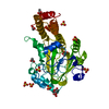


 Type: D-saccharide, beta linking / Mass: 180.156 Da / Num. of mol.: 2


 Mass: 96.063 Da / Num. of mol.: 7 / Source method: obtained synthetically / Formula: SO4
 Mass: 59.044 Da / Num. of mol.: 5 / Source method: obtained synthetically / Formula: C2H3O2
 Mass: 58.693 Da / Num. of mol.: 1 / Source method: obtained synthetically / Formula: Ni
 Sample preparation
 / Beamline: I02 / Wavelength: 0.9795
  Processing