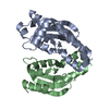
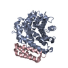
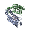
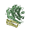
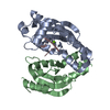
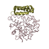
Entry Database : PDB / ID : 2goxTitle Crystal structure of Efb-C / C3d Complex Complement C3 Fibrinogen-binding protein Keywords / / Function / homology Function Domain/homology Component
/ / / / / / / / / / / / / / / / / / / / / / / / / / / / / / / / / / / / / / / / / / / / / / / / / / / / / / / / / / / / / / / / / / / / / / / / / / / / / / / / / / / / / / / / / / / / / / / / / / / / / / / / / / / / / / / / / / / / / / / / / / / / / / / Biological species Homo sapiens (human)Staphylococcus aureus subsp. aureus Mu50 (bacteria)Method / / / Resolution : 2.2 Å Authors Hammel, M. / Geisbrecht, B.V. Journal : Nat.Immunol. / Year : 2007Title : A structural basis for complement inhibition by Staphylococcus aureus.Authors : Hammel, M. / Sfyroera, G. / Ricklin, D. / Magotti, P. / Lambris, J.D. / Geisbrecht, B.V. History Deposition Apr 14, 2006 Deposition site / Processing site Revision 1.0 Mar 20, 2007 Provider / Type Revision 1.1 Sep 12, 2007 Group Revision 1.2 Jul 13, 2011 Group / Version format complianceRevision 1.3 Oct 18, 2017 Group / Category Revision 1.4 Oct 20, 2021 Group / Category / struct_ref_seq_difItem / _database_2.pdbx_database_accession / _struct_ref_seq_dif.detailsRevision 1.5 Aug 30, 2023 Group / Refinement descriptionCategory / chem_comp_bond / pdbx_initial_refinement_modelRevision 1.6 Nov 6, 2024 Group / Category / pdbx_modification_feature
Show all Show less
 Movie
Movie Controller


 Open data
 Basic information
 Components
 Keywords
 Function and homology information
 Homo sapiens (human)
 Staphylococcus aureus subsp. aureus Mu50 (bacteria)
 X-RAY DIFFRACTION /
  Authors
 Citation
 Structure visualization
 Downloads & links
 Other downloads


 PDBj
PDBj


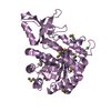
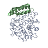
 Assembly
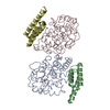
 Mass: 18.015 Da / Num. of mol.: 317 / Source method: isolated from a natural source / Formula: H2O
 Sample preparation
 / Beamline: 22-BM / Wavelength: 0.9184 Å
 Processing