 Movie
Movie Controller


+ Open data

- Basic information
| Entry | Database: PDB / ID: 3d5s | ||||||
|---|---|---|---|---|---|---|---|
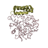
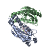
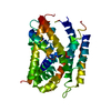
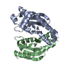
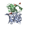
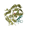
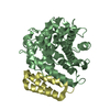
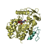
| Title | Crystal Structure of Efb-C (R131A) / C3d Complex | ||||||
 Components Components |
| ||||||
 Keywords Keywords | CELL ADHESION/TOXIN / Protein-protein complex / cell adhesion-toxin complex / site-directed mutation / Age-related macular degeneration / Cleavage on pair of basic residues / Complement alternate pathway / Complement pathway / Disease mutation / Glycoprotein / Immune response / Inflammatory response / Innate immunity / Phosphoprotein / Polymorphism / Secreted / Thioester bond | ||||||
| Function / homology |  Function and homology information Function and homology informationcomplement binding / C5L2 anaphylatoxin chemotactic receptor binding / oviduct epithelium development / regulation of triglyceride biosynthetic process / positive regulation of activation of membrane attack complex / vertebrate eye-specific patterning / positive regulation of apoptotic cell clearance / Alternative complement activation / complement-mediated synapse pruning / positive regulation of type IIa hypersensitivity ...complement binding / C5L2 anaphylatoxin chemotactic receptor binding / oviduct epithelium development / regulation of triglyceride biosynthetic process / positive regulation of activation of membrane attack complex / vertebrate eye-specific patterning / positive regulation of apoptotic cell clearance / Alternative complement activation / complement-mediated synapse pruning / positive regulation of type IIa hypersensitivity / Activation of C3 and C5 / positive regulation of lipid storage / complement activation, GZMK pathway / positive regulation of phagocytosis, engulfment / positive regulation of G protein-coupled receptor signaling pathway / complement-dependent cytotoxicity / positive regulation of D-glucose transmembrane transport / complement receptor mediated signaling pathway / complement activation, alternative pathway / complement activation / endopeptidase inhibitor activity / neuron remodeling / amyloid-beta clearance / B cell activation / complement activation, classical pathway / positive regulation of vascular endothelial growth factor production / Purinergic signaling in leishmaniasis infection / Regulation of Complement cascade / Peptide ligand-binding receptors / response to bacterium / Post-translational protein phosphorylation / fatty acid metabolic process / positive regulation of protein phosphorylation / positive regulation of receptor-mediated endocytosis / Regulation of Insulin-like Growth Factor (IGF) transport and uptake by Insulin-like Growth Factor Binding Proteins (IGFBPs) / Immunoregulatory interactions between a Lymphoid and a non-Lymphoid cell / positive regulation of angiogenesis / azurophil granule lumen / secretory granule lumen / blood microparticle / G alpha (i) signalling events / immune response / endoplasmic reticulum lumen / G protein-coupled receptor signaling pathway / inflammatory response / receptor ligand activity / signaling receptor binding / Neutrophil degranulation / cell surface / signal transduction / protein-containing complex / : / extracellular exosome / extracellular region / plasma membrane Similarity search - Function | ||||||
| Biological species |  Homo sapiens (human) Homo sapiens (human) Staphylococcus aureus subsp. aureus str. Newman (bacteria) Staphylococcus aureus subsp. aureus str. Newman (bacteria) | ||||||
| Method |  X-RAY DIFFRACTION / SYNCHROTRON / MOLECULAR REPLACEMENT / molecular replacement / Resolution: 2.3 Å X-RAY DIFFRACTION / SYNCHROTRON / MOLECULAR REPLACEMENT / molecular replacement / Resolution: 2.3 Å | ||||||
 Authors Authors | Geisbrecht, B.V. | ||||||
 Citation Citation | Journal: Protein Sci. / Year: 2008 Title: Electrostatic contributions drive the interaction between Staphylococcus aureus protein Efb-C and its complement target C3d. Authors: Haspel, N. / Ricklin, D. / Geisbrecht, B.V. / Kavraki, L.E. / Lambris, J.D. | ||||||
| History |
|
- Structure visualization
| Structure viewer | Molecule: MolmilJmol/JSmol |
|---|
- Downloads & links
-Download
| PDBx/mmCIF format | 3d5s.cif.gz | 151.6 KB | Display | PDBx/mmCIF format |
|---|---|---|---|---|
| PDB format | pdb3d5s.ent.gz | 119.9 KB | Display | PDB format |
| PDBx/mmJSON format | 3d5s.json.gz | Tree view | PDBx/mmJSON format | |
| Others |  Other downloads Other downloads |
-Validation report
| Arichive directory | https://data.pdbj.org/pub/pdb/validation_reports/d5/3d5sftp://data.pdbj.org/pub/pdb/validation_reports/d5/3d5s | HTTPS FTP |
|---|
-Related structure data
| Related structure data |  3d5rC 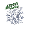 2goxS S: Starting model for refinement C: citing same article ( |
|---|---|
| Similar structure data |


-Links
 PDBj
PDBj


- Assembly
| Deposited unit | 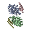
| ||||||||
|---|---|---|---|---|---|---|---|---|---|
| 1 | 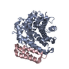
| ||||||||
| 2 | 
| ||||||||
| Unit cell |
|
-Components
| #1: Protein | Mass: 33143.879 Da / Num. of mol.: 2 / Fragment: Complement C3d fragment, UNP residues 996-1287 / Mutation: C1010A Source method: isolated from a genetically manipulated source Details: C3d coding sequence contains site-directed C1010A mutation Source: (gene. exp.) Homo sapiens (human) / Gene: C3, CPAMD1 / Production host: #2: Protein | Mass: 7506.786 Da / Num. of mol.: 2 / Fragment: C-terminal domain, UNP residues 101-165 / Mutation: R131A Source method: isolated from a genetically manipulated source Details: Efb-C coding seqeuence contains R131A site-directed mutation Source: (gene. exp.) Staphylococcus aureus subsp. aureus str. Newman (bacteria)Strain: Mu50 / Gene: fib, efb, fib, efb, NWMN_1069 / Production host: #3: Water | ChemComp-HOH / |  Mass: 18.015 Da / Num. of mol.: 140 / Source method: isolated from a natural source / Formula: H2O Mass: 18.015 Da / Num. of mol.: 140 / Source method: isolated from a natural source / Formula: H2OHas protein modification | Y | |
|---|
-Experimental details
-Experiment
| Experiment | Method: X-RAY DIFFRACTION / Number of used crystals: 1 |
|---|
- Sample preparation
| Crystal | Density Matthews: 3.12 Å3/Da / Density % sol: 60.58 % |
|---|---|
| Crystal grow | Temperature: 293 K / Method: vapor diffusion, hanging drop / pH: 7 Details: 60% (v/v) tacsimate, pH 7.0, VAPOR DIFFUSION, HANGING DROP, temperature 293K |
-Data collection
| Diffraction | Mean temperature: 93 K |
|---|---|
| Diffraction source | Source: SYNCHROTRON / Site: APS  / Beamline: 22-ID / Wavelength: 1 Å / Beamline: 22-ID / Wavelength: 1 Å |
| Detector | Type: MARMOSAIC 300 mm CCD / Detector: CCD / Date: Dec 14, 2006 |
| Radiation | Protocol: SINGLE WAVELENGTH / Monochromatic (M) / Laue (L): M / Scattering type: x-ray |
| Radiation wavelength | Wavelength: 1 Å / Relative weight: 1 |
| Reflection | Resolution: 2.3→50 Å / Num. obs: 45612 / % possible obs: 99.7 % / Redundancy: 3.8 % / Rmerge(I) obs: 0.101 / Χ2: 0.993 / Net I/σ(I): 11.7 |
| Reflection shell | Resolution: 2.3→2.38 Å / Redundancy: 3.6 % / Rmerge(I) obs: 0.413 / Num. unique all: 4490 / Χ2: 0.963 / % possible all: 98.9 |
-Phasing
| Phasing | Method: molecular replacement | |||||||||
|---|---|---|---|---|---|---|---|---|---|---|
| Phasing MR | Rfactor: 0.391 / Cor.coef. Fo:Fc: 0.685
|
- Processing
| Software |
| ||||||||||||||||||||||||||||||||
|---|---|---|---|---|---|---|---|---|---|---|---|---|---|---|---|---|---|---|---|---|---|---|---|---|---|---|---|---|---|---|---|---|---|
| Refinement | Method to determine structure: MOLECULAR REPLACEMENT Starting model: PDB entry 2GOX Resolution: 2.3→50 Å / FOM work R set: 0.847 / Cross valid method: THROUGHOUT
| ||||||||||||||||||||||||||||||||
| Displacement parameters | Biso mean: 42.44 Å2 | ||||||||||||||||||||||||||||||||
| Refinement step | Cycle: LAST / Resolution: 2.3→50 Å
| ||||||||||||||||||||||||||||||||
| Refine LS restraints |
|
