histone H3K36me/H3K36me2 demethylase activity / histone H3K9me/H3K9me2 demethylase activity / histone H3K27me2/H3K27me3 demethylase activity / negative regulation of rDNA heterochromatin formation / [histone H3]-dimethyl-L-lysine9 demethylase / histone H3K36 demethylase activity / histone H4K20 demethylase activity / Oxidoreductases; Acting on paired donors, with incorporation or reduction of molecular oxygen; With 2-oxoglutarate as one donor, and incorporation of one atom of oxygen into each donor / histone H3K4me3 reader activity / histone H3K9 demethylase activity ...histone H3K36me/H3K36me2 demethylase activity / histone H3K9me/H3K9me2 demethylase activity / histone H3K27me2/H3K27me3 demethylase activity / negative regulation of rDNA heterochromatin formation / [histone H3]-dimethyl-L-lysine9 demethylase / histone H3K36 demethylase activity / histone H4K20 demethylase activity / Oxidoreductases; Acting on paired donors, with incorporation or reduction of molecular oxygen; With 2-oxoglutarate as one donor, and incorporation of one atom of oxygen into each donor / histone H3K4me3 reader activity / histone H3K9 demethylase activity / positive regulation of transcription by RNA polymerase I / histone demethylase activity / Condensation of Prophase Chromosomes / transcription coregulator activity / G1/S transition of mitotic cell cycle / HDMs demethylate histones / brain development / nuclear membrane / chromatin remodeling / iron ion binding / chromatin binding / regulation of transcription by RNA polymerase II / positive regulation of DNA-templated transcription / nucleolus / positive regulation of transcription by RNA polymerase II / zinc ion binding / nucleoplasm / nucleus Similarity search - Function
Methane Monooxygenase Hydroxylase; Chain G, domain 1 - #1360 / Jumonji, helical domain / Jumonji helical domain / : / Cupin / JmjC domain, hydroxylase / A domain family that is part of the cupin metalloenzyme superfamily. / JmjC domain / JmjC domain profile. / Zinc finger, PHD-type, conserved site ...Methane Monooxygenase Hydroxylase; Chain G, domain 1 - #1360 / Jumonji, helical domain / Jumonji helical domain / : / Cupin / JmjC domain, hydroxylase / A domain family that is part of the cupin metalloenzyme superfamily. / JmjC domain / JmjC domain profile. / Zinc finger, PHD-type, conserved site / PHD-finger / Zinc finger PHD-type signature. / Methane Monooxygenase Hydroxylase; Chain G, domain 1 / Zinc finger PHD-type profile. / Zinc finger, PHD-finger / Zinc finger, PHD-type / PHD zinc finger / Zinc finger, FYVE/PHD-type / Jelly Rolls / Up-down Bundle / Sandwich / Mainly Beta / Mainly Alpha Similarity search - Domain/homology
Resolution: 2.55→107.29 Å / Cor.coef. Fo:Fc: 0.94 / Cor.coef. Fo:Fc free: 0.911 / SU B: 9.551 / SU ML: 0.208 / Cross valid method: THROUGHOUT / ESU R: 0.377 / ESU R Free: 0.28 / Stereochemistry target values: MAXIMUM LIKELIHOOD / Details: HYDROGENS HAVE BEEN ADDED IN THE RIDING POSITIONS
Rfactor
Num. reflection
% reflection
Selection details
Rfree
0.26481
1034
5.4 %
RANDOM
Rwork
0.20795
-
-
-
all
0.21092
18069
-
-
obs
0.21092
18069
99.98 %
-
Solvent computation
Ion probe radii: 0.8 Å / Shrinkage radii: 0.8 Å / VDW probe radii: 1.2 Å / Solvent model: MASK
Displacement parameters
Biso mean: 42.357 Å2
Refinement step
Cycle: LAST / Resolution: 2.55→107.29 Å
Protein
Nucleic acid
Ligand
Solvent
Total
Num. atoms
2875
0
62
37
2974
Refine LS restraints
Refine-ID
Type
Dev ideal
Dev ideal target
Number
X-RAY DIFFRACTION
r_bond_refined_d
0.013
0.02
3008
X-RAY DIFFRACTION
r_bond_other_d
0.002
0.02
1989
X-RAY DIFFRACTION
r_angle_refined_deg
1.573
1.963
4088
X-RAY DIFFRACTION
r_angle_other_deg
0.963
3.001
4842
X-RAY DIFFRACTION
r_dihedral_angle_1_deg
6.305
5
366
X-RAY DIFFRACTION
r_dihedral_angle_2_deg
36.151
23.926
135
X-RAY DIFFRACTION
r_dihedral_angle_3_deg
15.694
15
484
X-RAY DIFFRACTION
r_dihedral_angle_4_deg
16.614
15
16
X-RAY DIFFRACTION
r_chiral_restr
0.08
0.2
462
X-RAY DIFFRACTION
r_gen_planes_refined
0.006
0.021
3317
X-RAY DIFFRACTION
r_gen_planes_other
0.001
0.02
633
X-RAY DIFFRACTION
r_nbd_refined
X-RAY DIFFRACTION
r_nbd_other
X-RAY DIFFRACTION
r_nbtor_refined
X-RAY DIFFRACTION
r_nbtor_other
X-RAY DIFFRACTION
r_xyhbond_nbd_refined
X-RAY DIFFRACTION
r_xyhbond_nbd_other
X-RAY DIFFRACTION
r_metal_ion_refined
X-RAY DIFFRACTION
r_metal_ion_other
X-RAY DIFFRACTION
r_symmetry_vdw_refined
X-RAY DIFFRACTION
r_symmetry_vdw_other
X-RAY DIFFRACTION
r_symmetry_hbond_refined
X-RAY DIFFRACTION
r_symmetry_hbond_other
X-RAY DIFFRACTION
r_symmetry_metal_ion_refined
X-RAY DIFFRACTION
r_symmetry_metal_ion_other
X-RAY DIFFRACTION
r_mcbond_it
X-RAY DIFFRACTION
r_mcbond_other
X-RAY DIFFRACTION
r_mcangle_it
X-RAY DIFFRACTION
r_scbond_it
X-RAY DIFFRACTION
r_scangle_it
X-RAY DIFFRACTION
r_rigid_bond_restr
X-RAY DIFFRACTION
r_sphericity_free
X-RAY DIFFRACTION
r_sphericity_bonded
LS refinement shell
Resolution: 2.55→2.616 Å / Total num. of bins used: 20
Rfactor
Num. reflection
% reflection
Rfree
0.445
84
-
Rwork
0.321
1282
-
obs
-
-
100 %
+
About Yorodumi
-
News
-
Feb 9, 2022. New format data for meta-information of EMDB entries
New format data for meta-information of EMDB entries
Version 3 of the EMDB header file is now the official format.
The previous official version 1.9 will be removed from the archive.
In the structure databanks used in Yorodumi, some data are registered as the other names, "COVID-19 virus" and "2019-nCoV". Here are the details of the virus and the list of structure data.
Jan 31, 2019. EMDB accession codes are about to change! (news from PDBe EMDB page)
EMDB accession codes are about to change! (news from PDBe EMDB page)
The allocation of 4 digits for EMDB accession codes will soon come to an end. Whilst these codes will remain in use, new EMDB accession codes will include an additional digit and will expand incrementally as the available range of codes is exhausted. The current 4-digit format prefixed with “EMD-” (i.e. EMD-XXXX) will advance to a 5-digit format (i.e. EMD-XXXXX), and so on. It is currently estimated that the 4-digit codes will be depleted around Spring 2019, at which point the 5-digit format will come into force.
The EM Navigator/Yorodumi systems omit the EMD- prefix.
Related info.:Q: What is EMD? / ID/Accession-code notation in Yorodumi/EM Navigator
Yorodumi is a browser for structure data from EMDB, PDB, SASBDB, etc.
This page is also the successor to EM Navigator detail page, and also detail information page/front-end page for Omokage search.
The word "yorodu" (or yorozu) is an old Japanese word meaning "ten thousand". "mi" (miru) is to see.
Related info.:EMDB / PDB / SASBDB / Comparison of 3 databanks / Yorodumi Search / Aug 31, 2016. New EM Navigator & Yorodumi / Yorodumi Papers / Jmol/JSmol / Function and homology information / Changes in new EM Navigator and Yorodumi
 Movie
Movie Controller


 Open data
 Basic information
 Components
 Keywords
 Function and homology information
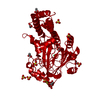
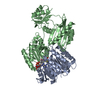
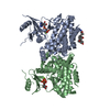
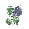
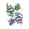
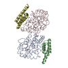
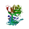
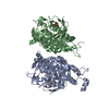
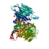
 Homo sapiens (human)
 X-RAY DIFFRACTION /
  Authors
 Structure visualization
 Downloads & links
 Other downloads

 PDBj
PDBj


 Assembly
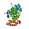


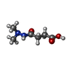


 Mass: 160.171 Da / Num. of mol.: 1 / Source method: obtained synthetically / Formula: C6H12N2O3
 Mass: 65.409 Da / Num. of mol.: 1 / Source method: obtained synthetically / Formula: Zn
 Mass: 96.063 Da / Num. of mol.: 6 / Source method: obtained synthetically / Formula: SO4
 Mass: 62.068 Da / Num. of mol.: 3 / Source method: obtained synthetically / Formula: C2H6O2
 Mass: 59.044 Da / Num. of mol.: 2 / Source method: obtained synthetically / Formula: C2H3O2
 Sample preparation
 / Beamline: I04-1 / Wavelength: 0.9173 Å
 Processing