 Movie
Movie Controller

[English] 日本語
 Yorodumi
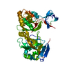
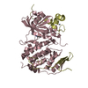
- PDB-1te1: Crystal structure of family 11 xylanase in complex with inhibitor... -
+ Open data

- Basic information
| Entry | Database: PDB / ID: 1te1 | ||||||
|---|---|---|---|---|---|---|---|
| Title | Crystal structure of family 11 xylanase in complex with inhibitor (XIP-I) | ||||||
 Components Components |
| ||||||
 Keywords Keywords | HYDROLASE INHIBITOR/HYDROLASE / BETA/ALPHA BARREL (XIP-I) and BETA JELLY ROLL (GH11) / HYDROLASE INHIBITOR-HYDROLASE COMPLEX | ||||||
| Function / homology |  Function and homology information Function and homology informationhydrolase activity, acting on glycosyl bonds / endo-1,4-beta-xylanase / endo-1,4-beta-xylanase activity / xylan catabolic process / defense response to fungus / enzyme inhibitor activity / carbohydrate metabolic process / extracellular region Similarity search - Function | ||||||
| Biological species |  Penicillium funiculosum (fungus) Penicillium funiculosum (fungus) | ||||||
| Method |  X-RAY DIFFRACTION / SYNCHROTRON / MOLECULAR REPLACEMENT / Resolution: 2.5 Å X-RAY DIFFRACTION / SYNCHROTRON / MOLECULAR REPLACEMENT / Resolution: 2.5 Å | ||||||
 Authors Authors | Payan, F. / Leone, P. / Furniss, C. / Tahir, T. / Durand, A. / Porciero, S. / Manzanares, P. / Williamson, G. / Gilbert, H.J. / Juge, N. / Roussel, A. | ||||||
 Citation Citation | Journal: J.Biol.Chem. / Year: 2004 Title: The Dual Nature of the Wheat Xylanase Protein Inhibitor XIP-I: STRUCTURAL BASIS FOR THE INHIBITION OF FAMILY 10 AND FAMILY 11 XYLANASES. Authors: Payan, F. / Leone, P. / Porciero, S. / Furniss, C. / Tahir, T. / Williamson, G. / Durand, A. / Manzanares, P. / Gilbert, H.J. / Juge, N. / Roussel, A. | ||||||
| History |
| ||||||
| Remark 999 | SEQUENCE THE AUTHORS BELIEVE THAT THESE RESIDUES ARE CORRECT AND GENEBANK IS INCORRECT AT THESE ...SEQUENCE THE AUTHORS BELIEVE THAT THESE RESIDUES ARE CORRECT AND GENEBANK IS INCORRECT AT THESE POSITIONS (SEE PDB ENTRY 1OM0). |
- Structure visualization
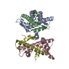
| Structure viewer | Molecule: MolmilJmol/JSmol |
|---|
- Downloads & links
-Download
| PDBx/mmCIF format | 1te1.cif.gz | 109 KB | Display | PDBx/mmCIF format |
|---|---|---|---|---|
| PDB format | pdb1te1.ent.gz | 82.8 KB | Display | PDB format |
| PDBx/mmJSON format | 1te1.json.gz | Tree view | PDBx/mmJSON format | |
| Others |  Other downloads Other downloads |
-Validation report
| Arichive directory | https://data.pdbj.org/pub/pdb/validation_reports/te/1te1ftp://data.pdbj.org/pub/pdb/validation_reports/te/1te1 | HTTPS FTP |
|---|
-Related structure data
| Related structure data |  1ta3C  1omoS  1ukrS S: Starting model for refinement C: citing same article ( |
|---|---|
| Similar structure data |


-Links
 PDBj
PDBj
- Assembly
| Deposited unit | 
| ||||||||
|---|---|---|---|---|---|---|---|---|---|
| 1 |
| ||||||||
| Unit cell |
|
-Components
| #1: Protein | Mass: 30323.990 Da / Num. of mol.: 1 / Source method: isolated from a natural source / Source: (natural) | ||||||
|---|---|---|---|---|---|---|---|
| #2: Protein | Mass: 20633.953 Da / Num. of mol.: 1 Source method: isolated from a genetically manipulated source Source: (gene. exp.) Penicillium funiculosum (fungus) / Gene: xynC / Production host: Penicillium funiculosum (fungus) / References: UniProt: Q9HFH0, endo-1,4-beta-xylanase | ||||||
| #3: Sugar |   Type: D-saccharide, beta linking / Mass: 221.208 Da / Num. of mol.: 2 Type: D-saccharide, beta linking / Mass: 221.208 Da / Num. of mol.: 2Source method: isolated from a genetically manipulated source Formula: C8H15NO6 #4: Chemical | ChemComp-EDO /   Mass: 62.068 Da / Num. of mol.: 11 / Source method: obtained synthetically / Formula: C2H6O2 Mass: 62.068 Da / Num. of mol.: 11 / Source method: obtained synthetically / Formula: C2H6O2#5: Water | ChemComp-HOH / |  Mass: 18.015 Da / Num. of mol.: 199 / Source method: isolated from a natural source / Formula: H2O Mass: 18.015 Da / Num. of mol.: 199 / Source method: isolated from a natural source / Formula: H2OHas protein modification | Y | |
-Experimental details
-Experiment
| Experiment | Method: X-RAY DIFFRACTION / Number of used crystals: 1 |
|---|
- Sample preparation
| Crystal | Density Matthews: 2.7 Å3/Da / Density % sol: 54 % |
|---|---|
| Crystal grow | Temperature: 293 K / Method: vapor diffusion, hanging drop / pH: 7 Details: PEG 4000, ammonium sulfate, 1,2,3-heptanetriol, pH 7.0, VAPOR DIFFUSION, HANGING DROP, temperature 293.0K |
-Data collection
| Diffraction | Mean temperature: 100 K |
|---|---|
| Diffraction source | Source: SYNCHROTRON / Site: SLS  / Beamline: X06SA / Wavelength: 0.9785 Å / Beamline: X06SA / Wavelength: 0.9785 Å |
| Detector | Type: MARRESEARCH / Detector: CCD / Date: Feb 12, 2004 |
| Radiation | Protocol: SINGLE WAVELENGTH / Monochromatic (M) / Laue (L): M / Scattering type: x-ray |
| Radiation wavelength | Wavelength: 0.9785 Å / Relative weight: 1 |
| Reflection | Resolution: 2.5→30 Å / Num. obs: 19734 / % possible obs: 99.8 % / Redundancy: 5.6 % / Biso Wilson estimate: 60.5 Å2 / Rmerge(I) obs: 0.075 / Rsym value: 0.068 / Net I/σ(I): 8.5 |
| Reflection shell | Resolution: 2.5→2.64 Å / Redundancy: 5.7 % / Rmerge(I) obs: 0.427 / Mean I/σ(I) obs: 1.9 / Num. unique all: 2834 / Rsym value: 0.387 / % possible all: 99.8 |
- Processing
| Software |
| ||||||||||||||||||||||||||||||||||||||||||||||||||||||||||||||||||||||||||||||||||||||||||||||||||||
|---|---|---|---|---|---|---|---|---|---|---|---|---|---|---|---|---|---|---|---|---|---|---|---|---|---|---|---|---|---|---|---|---|---|---|---|---|---|---|---|---|---|---|---|---|---|---|---|---|---|---|---|---|---|---|---|---|---|---|---|---|---|---|---|---|---|---|---|---|---|---|---|---|---|---|---|---|---|---|---|---|---|---|---|---|---|---|---|---|---|---|---|---|---|---|---|---|---|---|---|---|---|
| Refinement | Method to determine structure: MOLECULAR REPLACEMENT Starting model: PDB ENTRY 1UKR, PDB ENTRY 1OMO Resolution: 2.5→20 Å / Cor.coef. Fo:Fc: 0.944 / Cor.coef. Fo:Fc free: 0.903 / SU B: 11.999 / SU ML: 0.258 / Cross valid method: THROUGHOUT / ESU R: 0.635 / ESU R Free: 0.32 / Stereochemistry target values: MAXIMUM LIKELIHOOD / Details: HYDROGENS HAVE BEEN ADDED IN THE RIDING POSITIONS
| ||||||||||||||||||||||||||||||||||||||||||||||||||||||||||||||||||||||||||||||||||||||||||||||||||||
| Solvent computation | Ion probe radii: 0.8 Å / Shrinkage radii: 0.8 Å / VDW probe radii: 1.4 Å / Solvent model: BABINET MODEL WITH MASK | ||||||||||||||||||||||||||||||||||||||||||||||||||||||||||||||||||||||||||||||||||||||||||||||||||||
| Displacement parameters | Biso mean: 26.836 Å2
| ||||||||||||||||||||||||||||||||||||||||||||||||||||||||||||||||||||||||||||||||||||||||||||||||||||
| Refinement step | Cycle: LAST / Resolution: 2.5→20 Å
| ||||||||||||||||||||||||||||||||||||||||||||||||||||||||||||||||||||||||||||||||||||||||||||||||||||
| Refine LS restraints |
| ||||||||||||||||||||||||||||||||||||||||||||||||||||||||||||||||||||||||||||||||||||||||||||||||||||
| LS refinement shell | Resolution: 2.5→2.633 Å / Total num. of bins used: 10 /
| ||||||||||||||||||||||||||||||||||||||||||||||||||||||||||||||||||||||||||||||||||||||||||||||||||||
| Refinement TLS params. | Method: refined / Refine-ID: X-RAY DIFFRACTION
| ||||||||||||||||||||||||||||||||||||||||||||||||||||||||||||||||||||||||||||||||||||||||||||||||||||
| Refinement TLS group |
|
