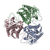
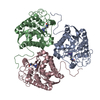
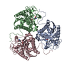
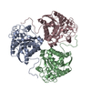
regulation of L-arginine import across plasma membrane / Urea cycle / collagen biosynthetic process / mammary gland involution / positive regulation of neutrophil mediated killing of fungus / negative regulation of T-helper 2 cell cytokine production / arginine metabolic process / arginase / arginase activity / response to selenium ion ...regulation of L-arginine import across plasma membrane / Urea cycle / collagen biosynthetic process / mammary gland involution / positive regulation of neutrophil mediated killing of fungus / negative regulation of T-helper 2 cell cytokine production / arginine metabolic process / arginase / arginase activity / response to selenium ion / urea cycle / response to nematode / response to methylmercury / response to manganese ion / response to steroid hormone / response to vitamin A / response to amine / response to herbicide / Neutrophil degranulation / response to zinc ion / response to vitamin E / defense response to protozoan / negative regulation of activated T cell proliferation / negative regulation of type II interferon-mediated signaling pathway / response to amino acid / maternal process involved in female pregnancy / cellular response to dexamethasone stimulus / response to cadmium ion / cellular response to transforming growth factor beta stimulus / response to axon injury / negative regulation of T cell proliferation / positive regulation of endothelial cell proliferation / cellular response to glucagon stimulus / lung development / cellular response to interleukin-4 / liver development / female pregnancy / response to peptide hormone / cellular response to hydrogen peroxide / manganese ion binding / cellular response to lipopolysaccharide / response to lipopolysaccharide / adaptive immune response / response to xenobiotic stimulus / innate immune response / neuronal cell body / : / identical protein binding / cytoplasm / cytosol Similarity search - Function
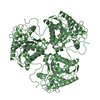
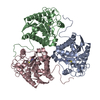
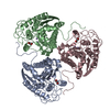
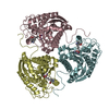
Arginase / Ureohydrolase domain / Ureohydrolase, manganese-binding site / Arginase family signature. / Ureohydrolase / Arginase family / Arginase family profile. / Arginase; Chain A / Ureohydrolase domain superfamily / 3-Layer(aba) Sandwich / Alpha Beta Similarity search - Domain/homology
In the structure databanks used in Yorodumi, some data are registered as the other names, "COVID-19 virus" and "2019-nCoV". Here are the details of the virus and the list of structure data.
Jan 31, 2019. EMDB accession codes are about to change! (news from PDBe EMDB page)
EMDB accession codes are about to change! (news from PDBe EMDB page)
The allocation of 4 digits for EMDB accession codes will soon come to an end. Whilst these codes will remain in use, new EMDB accession codes will include an additional digit and will expand incrementally as the available range of codes is exhausted. The current 4-digit format prefixed with “EMD-” (i.e. EMD-XXXX) will advance to a 5-digit format (i.e. EMD-XXXXX), and so on. It is currently estimated that the 4-digit codes will be depleted around Spring 2019, at which point the 5-digit format will come into force.
The EM Navigator/Yorodumi systems omit the EMD- prefix.
Related info.:Q: What is EMD? / ID/Accession-code notation in Yorodumi/EM Navigator
Yorodumi is a browser for structure data from EMDB, PDB, SASBDB, etc.
This page is also the successor to EM Navigator detail page, and also detail information page/front-end page for Omokage search.
The word "yorodu" (or yorozu) is an old Japanese word meaning "ten thousand". "mi" (miru) is to see.
Related info.:EMDB / PDB / SASBDB / Comparison of 3 databanks / Yorodumi Search / Aug 31, 2016. New EM Navigator & Yorodumi / Yorodumi Papers / Jmol/JSmol / Function and homology information / Changes in new EM Navigator and Yorodumi
 Movie
Movie Controller


 Open data
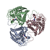
 Basic information
 Components
 Keywords
 Function and homology information
 X-RAY DIFFRACTION /
  Authors
 Citation
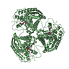
 Structure visualization
 Downloads & links
 Other downloads


 PDBj
PDBj


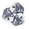
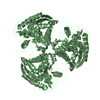
 Assembly
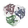

 Mass: 18.998 Da / Num. of mol.: 6 / Source method: obtained synthetically / Formula: F
 Mass: 54.938 Da / Num. of mol.: 6 / Source method: obtained synthetically / Formula: Mn
 Type: L-peptide linking / Mass: 175.209 Da / Num. of mol.: 3 / Source method: obtained synthetically / Formula: C6H15N4O2
 Mass: 18.015 Da / Num. of mol.: 100 / Source method: isolated from a natural source / Formula: H2O
 Sample preparation
 / Beamline: 5.0.1 / Wavelength: 0.982 Å
 Processing