 Movie
Movie Controller

[English] 日本語
 Yorodumi
- PDB-1t3m: Structure of the isoaspartyl peptidase with L-asparaginase activi... -
+ Open data

- Basic information
| Entry | Database: PDB / ID: 1t3m | ||||||
|---|---|---|---|---|---|---|---|
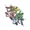
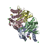
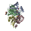
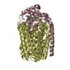
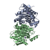
| Title | Structure of the isoaspartyl peptidase with L-asparaginase activity from E. coli | ||||||
 Components Components | (Putative L-asparaginase) x 2 | ||||||
 Keywords Keywords | HYDROLASE / TYPE III L-ASPARAGINASE / PLANT-TYPE ASPARAGINASE / ISOASPARTYL PEPTIDASE | ||||||
| Function / homology |  Function and homology information Function and homology informationbeta-aspartyl-peptidase / asparaginase activity / beta-aspartyl-peptidase activity / protein autoprocessing / hydrolase activity Similarity search - Function | ||||||
| Biological species |  | ||||||
| Method |  X-RAY DIFFRACTION / SYNCHROTRON / MOLECULAR REPLACEMENT / Resolution: 1.65 Å X-RAY DIFFRACTION / SYNCHROTRON / MOLECULAR REPLACEMENT / Resolution: 1.65 Å | ||||||
 Authors Authors | Prahl, A. / Pazgier, M. / Hejazi, M. / Lockau, W. / Lubkowski, J. | ||||||
 Citation Citation | Journal: Acta Crystallogr.,Sect.D / Year: 2004 Title: Structure of the isoaspartyl peptidase with L-asparaginase activity from Escherichia coli. Authors: Prahl, A. / Pazgier, M. / Hejazi, M. / Lockau, W. / Lubkowski, J. | ||||||
| History |
|
- Structure visualization
| Structure viewer | Molecule: MolmilJmol/JSmol |
|---|
- Downloads & links
-Download
| PDBx/mmCIF format | 1t3m.cif.gz | 140.3 KB | Display | PDBx/mmCIF format |
|---|---|---|---|---|
| PDB format | pdb1t3m.ent.gz | 106.5 KB | Display | PDB format |
| PDBx/mmJSON format | 1t3m.json.gz | Tree view | PDBx/mmJSON format | |
| Others |  Other downloads Other downloads |
-Validation report
| Summary document | 1t3m_validation.pdf.gz | 458.3 KB | Display | wwPDB validaton report |
|---|---|---|---|---|
| Full document | 1t3m_full_validation.pdf.gz | 464 KB | Display | |
| Data in XML | 1t3m_validation.xml.gz | 31.7 KB | Display | |
| Data in CIF | 1t3m_validation.cif.gz | 46.3 KB | Display | |
| Arichive directory | https://data.pdbj.org/pub/pdb/validation_reports/t3/1t3mftp://data.pdbj.org/pub/pdb/validation_reports/t3/1t3m | HTTPS FTP |
-Related structure data
| Related structure data |  1ayyS S: Starting model for refinement |
|---|---|
| Similar structure data |


-Links
 PDBj
PDBj


- Assembly
| Deposited unit | 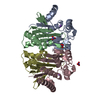
| ||||||||
|---|---|---|---|---|---|---|---|---|---|
| 1 |
| ||||||||
| Unit cell |
|
-Components
| #1: Protein | Mass: 18882.576 Da / Num. of mol.: 2 / Fragment: N-TERMINAL RESIDUES 1-177 Source method: isolated from a genetically manipulated source Source: (gene. exp.) #2: Protein | Mass: 14895.645 Da / Num. of mol.: 2 / Fragment: C-TERMINAL RESIDUES 178-320 Source method: isolated from a genetically manipulated source Source: (gene. exp.) #3: Chemical |   Mass: 22.990 Da / Num. of mol.: 2 / Source method: obtained synthetically / Formula: Na Mass: 22.990 Da / Num. of mol.: 2 / Source method: obtained synthetically / Formula: Na#4: Chemical |   Mass: 62.005 Da / Num. of mol.: 3 / Source method: obtained synthetically / Formula: NO3 Mass: 62.005 Da / Num. of mol.: 3 / Source method: obtained synthetically / Formula: NO3#5: Water | ChemComp-HOH / |  Mass: 18.015 Da / Num. of mol.: 681 / Source method: isolated from a natural source / Formula: H2O Mass: 18.015 Da / Num. of mol.: 681 / Source method: isolated from a natural source / Formula: H2O |
|---|
-Experimental details
-Experiment
| Experiment | Method: X-RAY DIFFRACTION / Number of used crystals: 1 |
|---|
- Sample preparation
| Crystal | Density Matthews: 2.63 Å3/Da / Density % sol: 53.21 % |
|---|---|
| Crystal grow | Temperature: 295 K / Method: vapor diffusion, hanging drop / pH: 7.5 Details: 15% PEG4000, 15% GLYCEROL, 0.3 M MAGNESIUM NITRATE, 0.1 M BIS-TRIS-HCL, pH 7.5, VAPOR DIFFUSION, HANGING DROP, temperature 295K |
-Data collection
| Diffraction | Mean temperature: 100 K |
|---|---|
| Diffraction source | Source: SYNCHROTRON / Site: APS  / Beamline: 22-ID / Wavelength: 0.98 / Wavelength: 0.98 Å / Beamline: 22-ID / Wavelength: 0.98 / Wavelength: 0.98 Å |
| Detector | Type: MAR CCD 165 mm / Detector: CCD |
| Radiation | Protocol: SINGLE WAVELENGTH / Monochromatic (M) / Laue (L): M / Scattering type: x-ray |
| Radiation wavelength | Wavelength: 0.98 Å / Relative weight: 1 |
| Reflection | Resolution: 1.65→30 Å / Num. obs: 84176 / % possible obs: 98.9 % / Observed criterion σ(F): -3 / Observed criterion σ(I): -3 / Redundancy: 4.7 % / Rmerge(I) obs: 0.01 / Net I/σ(I): 12.1 |
| Reflection shell | Resolution: 1.65→1.71 Å / Redundancy: 3.1 % / Rmerge(I) obs: 0.528 / Mean I/σ(I) obs: 1.97 / % possible all: 97.2 |
- Processing
| Software |
| |||||||||||||||||||||||||||||||||
|---|---|---|---|---|---|---|---|---|---|---|---|---|---|---|---|---|---|---|---|---|---|---|---|---|---|---|---|---|---|---|---|---|---|---|
| Refinement | Method to determine structure: MOLECULAR REPLACEMENT Starting model: PDB ENTRY 1AYY Resolution: 1.65→10 Å / Num. parameters: 20273 / Num. restraintsaints: 17952 / Cross valid method: FREE R / σ(F): 0 / Stereochemistry target values: ENGH AND HUBER Details: ANISOTROPIC SCALING APPLIED BY THE METHOD OF PARKIN, MOEZZI & HOPE, J.APPL.CRYST.28(1995)53-56. ANISOTROPIC REFINEMENT REDUCED FREE R (NO CUTOFF) BY 0.024.
| |||||||||||||||||||||||||||||||||
| Refine analyze | Num. disordered residues: 21 / Occupancy sum hydrogen: 0 / Occupancy sum non hydrogen: 4852.6 | |||||||||||||||||||||||||||||||||
| Refinement step | Cycle: LAST / Resolution: 1.65→10 Å
| |||||||||||||||||||||||||||||||||
| Refine LS restraints |
|
