 Movie
Movie Controller


+ Open data

- Basic information
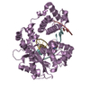
| Entry | Database: PDB / ID: 1ayy | ||||||
|---|---|---|---|---|---|---|---|
| Title | GLYCOSYLASPARAGINASE | ||||||
 Components Components | (GLYCOSYLASPARAGINASE) x 2 | ||||||
 Keywords Keywords | HYDROLASE / GLYCOAMIDASE | ||||||
| Function / homology |  Function and homology information Function and homology informationN4-(beta-N-acetylglucosaminyl)-L-asparaginase / N4-(beta-N-acetylglucosaminyl)-L-asparaginase activity / peptidase activity / periplasmic space / proteolysis / cytoplasm Similarity search - Function | ||||||
| Biological species |  Elizabethkingia meningoseptica (bacteria) Elizabethkingia meningoseptica (bacteria) | ||||||
| Method |  X-RAY DIFFRACTION / MOLECULAR REPLACEMENT, PHASE COMBINATION WITH 2 DERIVATIVES / Resolution: 2.32 Å X-RAY DIFFRACTION / MOLECULAR REPLACEMENT, PHASE COMBINATION WITH 2 DERIVATIVES / Resolution: 2.32 Å | ||||||
 Authors Authors | Van Roey, P. / Xuan, J. | ||||||
 Citation Citation | Journal: Protein Sci. / Year: 1998 Title: Crystal structure of glycosylasparaginase from Flavobacterium meningosepticum. Authors: Xuan, J. / Tarentino, A.L. / Grimwood, B.G. / Plummer Jr., T.H. / Cui, T. / Guan, C. / Van Roey, P. | ||||||
| History |
|
- Structure visualization
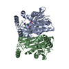
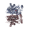
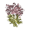
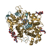
| Structure viewer | Molecule: MolmilJmol/JSmol |
|---|
- Downloads & links
-Download
| PDBx/mmCIF format | 1ayy.cif.gz | 119.6 KB | Display | PDBx/mmCIF format |
|---|---|---|---|---|
| PDB format | pdb1ayy.ent.gz | 93 KB | Display | PDB format |
| PDBx/mmJSON format | 1ayy.json.gz | Tree view | PDBx/mmJSON format | |
| Others |  Other downloads Other downloads |
-Validation report
| Arichive directory | https://data.pdbj.org/pub/pdb/validation_reports/ay/1ayyftp://data.pdbj.org/pub/pdb/validation_reports/ay/1ayy | HTTPS FTP |
|---|
-Related structure data
| Similar structure data |
|---|


-Links
 PDBj
PDBj


- Assembly
| Deposited unit | 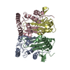
| ||||||||
|---|---|---|---|---|---|---|---|---|---|
| 1 |
| ||||||||
| Unit cell |
|
-Components
| #1: Protein | Mass: 16819.072 Da / Num. of mol.: 2 Source method: isolated from a genetically manipulated source Source: (gene. exp.) Elizabethkingia meningoseptica (bacteria)Production host: References: UniProt: Q47898, N4-(beta-N-acetylglucosaminyl)-L-asparaginase #2: Protein | Mass: 15378.576 Da / Num. of mol.: 2 Source method: isolated from a genetically manipulated source Source: (gene. exp.) Elizabethkingia meningoseptica (bacteria)Production host: References: UniProt: Q47898, N4-(beta-N-acetylglucosaminyl)-L-asparaginase #3: Water | ChemComp-HOH / |  Mass: 18.015 Da / Num. of mol.: 200 / Source method: isolated from a natural source / Formula: H2O Mass: 18.015 Da / Num. of mol.: 200 / Source method: isolated from a natural source / Formula: H2OCompound details | THERE IS A SINGLE CHAIN PRECURSOR ACTIVATED BY PROTEOLYTI | |
|---|
-Experimental details
-Experiment
| Experiment | Method: X-RAY DIFFRACTION / Number of used crystals: 1 |
|---|
- Sample preparation
| Crystal | Density Matthews: 2.1 Å3/Da / Density % sol: 41 % | ||||||||||||||||||||||||||||||||||||||||||
|---|---|---|---|---|---|---|---|---|---|---|---|---|---|---|---|---|---|---|---|---|---|---|---|---|---|---|---|---|---|---|---|---|---|---|---|---|---|---|---|---|---|---|---|
| Crystal grow | pH: 8.5 / Details: 19% PEG3350 IN 100 MM TRIS.HCL PH 8.5 | ||||||||||||||||||||||||||||||||||||||||||
| Crystal grow | *PLUS Temperature: 10 ℃ / pH: 7.5 / Method: vapor diffusion, hanging drop | ||||||||||||||||||||||||||||||||||||||||||
| Components of the solutions | *PLUS
|
-Data collection
| Diffraction | Mean temperature: 130 K |
|---|---|
| Diffraction source | Source: ROTATING ANODE / Type: RIGAKU RUH2R / Wavelength: 1.5418 |
| Detector | Type: RIGAKU / Detector: IMAGE PLATE / Date: Nov 1, 1996 / Details: MIRRORS |
| Radiation | Monochromator: NI FILTER / Monochromatic (M) / Laue (L): M / Scattering type: x-ray |
| Radiation wavelength | Wavelength: 1.5418 Å / Relative weight: 1 |
| Reflection | Resolution: 2.32→20 Å / Num. obs: 22627 / % possible obs: 95.7 % / Observed criterion σ(I): 0 / Redundancy: 2.9 % / Rmerge(I) obs: 0.075 / Rsym value: 0.075 / Net I/σ(I): 7 |
| Reflection shell | Resolution: 2.32→2.4 Å / Redundancy: 2.73 % / Rmerge(I) obs: 0.17 / Mean I/σ(I) obs: 3 / Rsym value: 0.17 / % possible all: 72.2 |
- Processing
| Software |
| ||||||||||||||||||||||||||||||||||||||||||||||||||||||||||||
|---|---|---|---|---|---|---|---|---|---|---|---|---|---|---|---|---|---|---|---|---|---|---|---|---|---|---|---|---|---|---|---|---|---|---|---|---|---|---|---|---|---|---|---|---|---|---|---|---|---|---|---|---|---|---|---|---|---|---|---|---|---|
| Refinement | Method to determine structure: MOLECULAR REPLACEMENT, PHASE COMBINATION WITH 2 DERIVATIVES Starting model: HUMAN GLYCOSYLASPARAGINASE DIMER Resolution: 2.32→20 Å / Data cutoff low absF: 0 / Cross valid method: THROUGHOUT / σ(F): 0
| ||||||||||||||||||||||||||||||||||||||||||||||||||||||||||||
| Displacement parameters | Biso mean: 26.5 Å2 | ||||||||||||||||||||||||||||||||||||||||||||||||||||||||||||
| Refinement step | Cycle: LAST / Resolution: 2.32→20 Å
| ||||||||||||||||||||||||||||||||||||||||||||||||||||||||||||
| Refine LS restraints |
| ||||||||||||||||||||||||||||||||||||||||||||||||||||||||||||
| LS refinement shell | Resolution: 2.32→2.4 Å / Total num. of bins used: 8
| ||||||||||||||||||||||||||||||||||||||||||||||||||||||||||||
| Xplor file | Serial no: 1 / Param file: PARAM19X.PRO / Topol file: TOPH19X.PRO | ||||||||||||||||||||||||||||||||||||||||||||||||||||||||||||
| Software | *PLUS Name: X-PLOR / Version: 3.8 / Classification: refinement | ||||||||||||||||||||||||||||||||||||||||||||||||||||||||||||
| Refinement | *PLUS Rfactor Rfree: 0.27 | ||||||||||||||||||||||||||||||||||||||||||||||||||||||||||||
| Solvent computation | *PLUS | ||||||||||||||||||||||||||||||||||||||||||||||||||||||||||||
| Displacement parameters | *PLUS | ||||||||||||||||||||||||||||||||||||||||||||||||||||||||||||
| Refine LS restraints | *PLUS
|
