Monochromator: SI 111 CHANNEL / Protocol: SINGLE WAVELENGTH / Monochromatic (M) / Laue (L): M / Scattering type: x-ray
Radiation wavelength
Wavelength: 0.97915 Å / Relative weight: 1
Reflection
Resolution: 3→99.9 Å / Num. obs: 67235 / % possible obs: 99.9 % / Observed criterion σ(I): 2.39 / Redundancy: 4.98 % / Biso Wilson estimate: 41.81 Å2 / Rmerge(I) obs: 0.1046 / Net I/σ(I): 13.34
Reflection shell
Resolution: 3.1→3.2 Å / Redundancy: 5.02 % / Rmerge(I) obs: 0.47 / Mean I/σ(I) obs: 2.86 / % possible all: 99.9
-
Processing
Software
Name
Version
Classification
ADSC
Quantum
datacollection
SHARP
phasing
REFMAC
5.6.0117
refinement
XDS
datareduction
XDS
datascaling
BUSTER
2.10.0
refinement
Refinement
Method to determine structure: SAD / Resolution: 3→34.64 Å / Cor.coef. Fo:Fc: 0.8414 / Cor.coef. Fo:Fc free: 0.826 / SU B: 62.315 / SU ML: 0.488 / Cross valid method: THROUGHOUT / σ(F): 0 / ESU R Free: 0.496 / Stereochemistry target values: MAXIMUM LIKELIHOOD / Details: HYDROGENS HAVE BEEN ADDED IN THE RIDING POSITIONS
Rfactor
Num. reflection
% reflection
Selection details
Rfree
0.2318
3401
5.06 %
RANDOM
Rwork
0.198
-
-
-
obs
0.1997
67174
99.94 %
-
Solvent computation
Ion probe radii: 0.8 Å / Shrinkage radii: 0.8 Å / VDW probe radii: 1.2 Å / Solvent model: MASK
Displacement parameters
Biso mean: 84.68 Å2
Baniso -1
Baniso -2
Baniso -3
1-
-13.5174 Å2
0 Å2
29.4009 Å2
2-
-
-12.1713 Å2
0 Å2
3-
-
-
25.6887 Å2
Refine analyze
Luzzati coordinate error obs: 0.448 Å
Refinement step
Cycle: LAST / Resolution: 3→34.64 Å
Protein
Nucleic acid
Ligand
Solvent
Total
Num. atoms
18425
0
1385
0
19810
Refine LS restraints
Refine-ID
Type
Dev ideal
Number
Restraint function
Weight
X-RAY DIFFRACTION
t_bond_d
0.01
20405
HARMONIC
2
X-RAY DIFFRACTION
t_angle_deg
1.24
27989
HARMONIC
2
X-RAY DIFFRACTION
t_dihedral_angle_d
7056
SINUSOIDAL
2
X-RAY DIFFRACTION
t_incorr_chiral_ct
X-RAY DIFFRACTION
t_pseud_angle
X-RAY DIFFRACTION
t_trig_c_planes
486
HARMONIC
2
X-RAY DIFFRACTION
t_gen_planes
2903
HARMONIC
5
X-RAY DIFFRACTION
t_it
20405
HARMONIC
20
X-RAY DIFFRACTION
t_nbd
X-RAY DIFFRACTION
t_omega_torsion
X-RAY DIFFRACTION
t_other_torsion
X-RAY DIFFRACTION
t_improper_torsion
X-RAY DIFFRACTION
t_chiral_improper_torsion
X-RAY DIFFRACTION
t_sum_occupancies
X-RAY DIFFRACTION
t_utility_distance
X-RAY DIFFRACTION
t_utility_angle
X-RAY DIFFRACTION
t_utility_torsion
X-RAY DIFFRACTION
t_ideal_dist_contact
LS refinement shell
Resolution: 3→3.08 Å / Total num. of bins used: 20
Rfactor
Num. reflection
% reflection
Rfree
0.3006
250
5.1 %
Rwork
0.2653
4653
-
all
0.2671
4903
-
obs
-
-
99.94 %
Refinement TLS params.
Method: refined / Refine-ID: X-RAY DIFFRACTION
ID
L11 (°2)
L12 (°2)
L13 (°2)
L22 (°2)
L23 (°2)
L33 (°2)
S11 (Å °)
S12 (Å °)
S13 (Å °)
S21 (Å °)
S22 (Å °)
S23 (Å °)
S31 (Å °)
S32 (Å °)
S33 (Å °)
T11 (Å2)
T12 (Å2)
T13 (Å2)
T22 (Å2)
T23 (Å2)
T33 (Å2)
Origin x (Å)
Origin y (Å)
Origin z (Å)
1
2.5757
0.1293
-1.2931
1.3256
0.3587
1.8121
-0.3427
-0.0441
-0.4348
0.0975
0.124
0.2484
0.3012
0.3239
0.2187
-0.1098
0.201
0.4262
-0.2053
0.1051
0.3851
-6.9674
-20.1968
-45.2696
2
5.3522
-2.1626
-1.5481
2.5382
0.705
1.371
0.0368
0.1854
0.1059
-0.0751
-0.0133
-0.3153
0.0348
-0.1434
-0.0235
-0.2228
0.0466
0.5003
-0.283
0.026
0.4651
9.121
-46.1524
-7.3396
3
23.8756
3.3756
-8.4438
2.1618
-1.8217
4.3857
-0.6535
0.5899
-1.9846
-0.1089
0.0776
-0.364
0.1697
-0.2244
0.5759
-0.8599
-0.0062
0.5755
-0.8462
0.05
0.1584
70.5065
-104.298
-71.949
4
2.0122
-0.3363
-0.6702
3.1157
-0.4474
0.7729
0.2487
0.0949
0.7089
0.0397
-0.0117
-0.2804
-0.1977
0.1071
-0.2369
-0.3073
-0.027
0.4508
-0.0013
0.0787
0.4066
14.4019
-88.2253
-53.8675
5
2.3718
0.391
-1.1021
2.9112
-0.8941
1.105
-0.2962
0.0318
-0.5875
-0.2601
-0.0337
-0.3091
0.2533
0.0855
0.3299
-0.1531
-0.0537
0.4188
-0.2198
-0.1569
0.2986
-43.5828
-62.1747
20.0266
6
3.2556
0.5052
-1.0953
1.3172
-0.398
1.4423
0.2123
-0.0216
0.6561
-0.1329
0.0993
-0.1812
-0.2403
0.0516
-0.3116
-0.118
0.0832
0.5866
-0.398
-0.0203
0.6538
-38.5896
-72.3979
-32.4114
Refinement TLS group
ID
Refine-ID
Refine TLS-ID
Selection details
Auth asym-ID
Auth seq-ID
1
X-RAY DIFFRACTION
1
{ A|37 - A|429 A|701 - A|722 }
A
37 - 429
2
X-RAY DIFFRACTION
1
{ A|37 - A|429 A|701 - A|722 }
A
701 - 722
3
X-RAY DIFFRACTION
2
{ B|36 - B|428 B|701 - B|716 }
B
36 - 428
4
X-RAY DIFFRACTION
2
{ B|36 - B|428 B|701 - B|716 }
B
701 - 716
5
X-RAY DIFFRACTION
3
{ C|48 - C|429 C|701 - C|712 }
C
48 - 429
6
X-RAY DIFFRACTION
3
{ C|48 - C|429 C|701 - C|712 }
C
701 - 712
7
X-RAY DIFFRACTION
4
{ D|39 - D|428 D|701 - D|716 }
D
39 - 428
8
X-RAY DIFFRACTION
4
{ D|39 - D|428 D|701 - D|716 }
D
701 - 716
9
X-RAY DIFFRACTION
5
{ E|38 - E|427 E|701 - E|721 }
E
38 - 427
10
X-RAY DIFFRACTION
5
{ E|38 - E|427 E|701 - E|721 }
E
701 - 721
11
X-RAY DIFFRACTION
6
{ F|40 - F|429 F|701 - F|723 }
F
40 - 429
12
X-RAY DIFFRACTION
6
{ F|40 - F|429 F|701 - F|723 }
F
701 - 723
+
About Yorodumi
-
News
-
Feb 9, 2022. New format data for meta-information of EMDB entries
New format data for meta-information of EMDB entries
Version 3 of the EMDB header file is now the official format.
The previous official version 1.9 will be removed from the archive.
In the structure databanks used in Yorodumi, some data are registered as the other names, "COVID-19 virus" and "2019-nCoV". Here are the details of the virus and the list of structure data.
Jan 31, 2019. EMDB accession codes are about to change! (news from PDBe EMDB page)
EMDB accession codes are about to change! (news from PDBe EMDB page)
The allocation of 4 digits for EMDB accession codes will soon come to an end. Whilst these codes will remain in use, new EMDB accession codes will include an additional digit and will expand incrementally as the available range of codes is exhausted. The current 4-digit format prefixed with “EMD-” (i.e. EMD-XXXX) will advance to a 5-digit format (i.e. EMD-XXXXX), and so on. It is currently estimated that the 4-digit codes will be depleted around Spring 2019, at which point the 5-digit format will come into force.
The EM Navigator/Yorodumi systems omit the EMD- prefix.
Related info.:Q: What is EMD? / ID/Accession-code notation in Yorodumi/EM Navigator
Yorodumi is a browser for structure data from EMDB, PDB, SASBDB, etc.
This page is also the successor to EM Navigator detail page, and also detail information page/front-end page for Omokage search.
The word "yorodu" (or yorozu) is an old Japanese word meaning "ten thousand". "mi" (miru) is to see.
Related info.:EMDB / PDB / SASBDB / Comparison of 3 databanks / Yorodumi Search / Aug 31, 2016. New EM Navigator & Yorodumi / Yorodumi Papers / Jmol/JSmol / Function and homology information / Changes in new EM Navigator and Yorodumi
 Movie
Movie Controller


 Open data
 Basic information
 Components
 Keywords
 Function and homology information
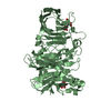
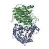
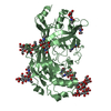
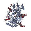
 Homo sapiens (human)
 X-RAY DIFFRACTION /
  Authors
 Citation
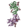
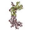
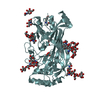
 Structure visualization
 Downloads & links
 Other downloads
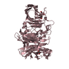


 PDBj
PDBj Assembly
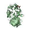
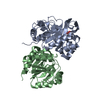
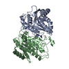

 Type: D-saccharide, beta linking / Mass: 221.208 Da / Num. of mol.: 15

 Mass: 94.971 Da / Num. of mol.: 6 / Source method: obtained synthetically / Formula: PO4
 Mass: 106.120 Da / Num. of mol.: 2 / Source method: obtained synthetically / Formula: C4H10O3
 Sample preparation
 / Beamline: 19-ID / Wavelength: 0.97915
  Processing