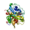
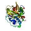
Trafficking and processing of endosomal TLR / Collagen degradation / cathepsin B / MHC class II antigen presentation / Neutrophil degranulation / proteolysis involved in protein catabolic process / melanosome / endopeptidase activity / lysosome / apical plasma membrane ...Trafficking and processing of endosomal TLR / Collagen degradation / cathepsin B / MHC class II antigen presentation / Neutrophil degranulation / proteolysis involved in protein catabolic process / melanosome / endopeptidase activity / lysosome / apical plasma membrane / cysteine-type endopeptidase activity / extracellular space Similarity search - Function
Peptidase C1A, propeptide / Peptidase family C1 propeptide / Single alpha-helices involved in coiled-coils or other helix-helix interfaces - #170 / Cysteine peptidase, asparagine active site / Eukaryotic thiol (cysteine) proteases asparagine active site. / Cysteine peptidase, histidine active site / Eukaryotic thiol (cysteine) proteases histidine active site. / : / Peptidase C1A, papain C-terminal / Papain family cysteine protease ...Peptidase C1A, propeptide / Peptidase family C1 propeptide / Single alpha-helices involved in coiled-coils or other helix-helix interfaces - #170 / Cysteine peptidase, asparagine active site / Eukaryotic thiol (cysteine) proteases asparagine active site. / Cysteine peptidase, histidine active site / Eukaryotic thiol (cysteine) proteases histidine active site. / : / Peptidase C1A, papain C-terminal / Papain family cysteine protease / Papain family cysteine protease / Cysteine proteinases / Cysteine peptidase, cysteine active site / Eukaryotic thiol (cysteine) proteases cysteine active site. / Cathepsin B; Chain A / Single alpha-helices involved in coiled-coils or other helix-helix interfaces / Papain-like cysteine peptidase superfamily / Alpha-Beta Complex / Up-down Bundle / Mainly Alpha / Alpha Beta Similarity search - Domain/homology
SEQUENCE THE PROTEIN IS POSTRANSLATIONALLY MODIFIED. AFTER THE ENZYME IS PROCESSED, THE PEPTIDE ...SEQUENCE THE PROTEIN IS POSTRANSLATIONALLY MODIFIED. AFTER THE ENZYME IS PROCESSED, THE PEPTIDE BOND BETWEEN THE RESIDUES 48 49 IS CLEAVED.
Type: peptide-like, Peptide-like / Class: Inhibitor / Mass: 583.631 Da / Num. of mol.: 1 / Source method: obtained synthetically / Formula: C26H41N5O10 Details: LEU-GLY-MEU. EPO is linked to the LEU-GLY-MEU as well as LEU-PRO. The inhibitor was chemically synthesized. References: NS-134 TWO HEADED EPOXYSUCCINYL INHIBITOR
Mass: 18.015 Da / Num. of mol.: 176 / Source method: isolated from a natural source / Formula: H2O
Has protein modification
Y
Nonpolymer details
THE INHIBITOR IS A TWO HEADED EPOXYSUCCINYL INHIBITOR. THE EPOXY GROUP OPENS UPON REACTION WITH THE ...THE INHIBITOR IS A TWO HEADED EPOXYSUCCINYL INHIBITOR. THE EPOXY GROUP OPENS UPON REACTION WITH THE ENZYME. THE INHIBITOR HAS TWO C-TERMINI AND NO N TERMINUS.
-
Experimental details
-
Experiment
Experiment
Method: X-RAY DIFFRACTION / Number of used crystals: 1
-
Sample preparation
Crystal
Density Matthews: 3.41 Å3/Da / Density % sol: 63.89 %
In the structure databanks used in Yorodumi, some data are registered as the other names, "COVID-19 virus" and "2019-nCoV". Here are the details of the virus and the list of structure data.
Jan 31, 2019. EMDB accession codes are about to change! (news from PDBe EMDB page)
EMDB accession codes are about to change! (news from PDBe EMDB page)
The allocation of 4 digits for EMDB accession codes will soon come to an end. Whilst these codes will remain in use, new EMDB accession codes will include an additional digit and will expand incrementally as the available range of codes is exhausted. The current 4-digit format prefixed with “EMD-” (i.e. EMD-XXXX) will advance to a 5-digit format (i.e. EMD-XXXXX), and so on. It is currently estimated that the 4-digit codes will be depleted around Spring 2019, at which point the 5-digit format will come into force.
The EM Navigator/Yorodumi systems omit the EMD- prefix.
Related info.:Q: What is EMD? / ID/Accession-code notation in Yorodumi/EM Navigator
Yorodumi is a browser for structure data from EMDB, PDB, SASBDB, etc.
This page is also the successor to EM Navigator detail page, and also detail information page/front-end page for Omokage search.
The word "yorodu" (or yorozu) is an old Japanese word meaning "ten thousand". "mi" (miru) is to see.
Related info.:EMDB / PDB / SASBDB / Comparison of 3 databanks / Yorodumi Search / Aug 31, 2016. New EM Navigator & Yorodumi / Yorodumi Papers / Jmol/JSmol / Function and homology information / Changes in new EM Navigator and Yorodumi
 Movie
Movie Controller

 Yorodumi
 Open data
 Basic information
 Components
 Keywords
 Function and homology information
 X-RAY DIFFRACTION /
  Authors
 Citation
 Structure visualization
 Downloads & links
 Other downloads


 PDBj
PDBj
 Assembly

 Type: peptide-like, Peptide-like / Class: Inhibitor / Mass: 583.631 Da / Num. of mol.: 1 / Source method: obtained synthetically / Formula: C26H41N5O10
 Mass: 18.015 Da / Num. of mol.: 176 / Source method: isolated from a natural source / Formula: H2O
 Sample preparation
 Processing