 Movie
Movie Controller

[English] 日本語
 Yorodumi
- PDB-2dca: X-ray crystal structure analysis of bovine spleen cathepsin B-CA0... -
+ Open data

- Basic information
| Entry | Database: PDB / ID: 2dca | ||||||
|---|---|---|---|---|---|---|---|
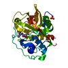
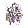
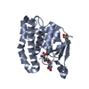
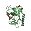
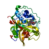
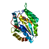
| Title | X-ray crystal structure analysis of bovine spleen cathepsin B-CA075 complex | ||||||
 Components Components | CATHEPSIN B | ||||||
 Keywords Keywords | HYDROLASE / Cathepsin B / Cysteine Protease / CA075 / EC 3.4.22.1 | ||||||
| Function / homology |  Function and homology information Function and homology informationTrafficking and processing of endosomal TLR / Collagen degradation / cathepsin B / MHC class II antigen presentation / Neutrophil degranulation / : / melanosome / endopeptidase activity / lysosome / apical plasma membrane ...Trafficking and processing of endosomal TLR / Collagen degradation / cathepsin B / MHC class II antigen presentation / Neutrophil degranulation / : / melanosome / endopeptidase activity / lysosome / apical plasma membrane / cysteine-type endopeptidase activity / : Similarity search - Function | ||||||
| Biological species |  | ||||||
| Method |  X-RAY DIFFRACTION / ISOMORPHOUS REPLACEMENT / Resolution: 2.11 Å X-RAY DIFFRACTION / ISOMORPHOUS REPLACEMENT / Resolution: 2.11 Å | ||||||
 Authors Authors | Watanabe, D. | ||||||
 Citation Citation | Journal: To be Published Title: Quantitative estimation of each active subsite of cathepsin B for the inhibitory activity, based on the inhibitory activitybinding mode relationship of a series of epoxysuccinyl inhibitors by ...Title: Quantitative estimation of each active subsite of cathepsin B for the inhibitory activity, based on the inhibitory activitybinding mode relationship of a series of epoxysuccinyl inhibitors by X-ray crystal structure analyses of the complexes Authors: Watanabe, D. / Yamamoto, A. / Matsumoto, K. / Murata, M. / Kitamura, K. / Tomoo, K. / Ishida, T. | ||||||
| History |
|
- Structure visualization
| Structure viewer | Molecule: MolmilJmol/JSmol |
|---|
- Downloads & links
-Download
| PDBx/mmCIF format | 2dca.cif.gz | 66.7 KB | Display | PDBx/mmCIF format |
|---|---|---|---|---|
| PDB format | pdb2dca.ent.gz | 48 KB | Display | PDB format |
| PDBx/mmJSON format | 2dca.json.gz | Tree view | PDBx/mmJSON format | |
| Others |  Other downloads Other downloads |
-Validation report
| Arichive directory | https://data.pdbj.org/pub/pdb/validation_reports/dc/2dcaftp://data.pdbj.org/pub/pdb/validation_reports/dc/2dca | HTTPS FTP |
|---|
-Related structure data
| Related structure data |  2dc6C  2dc7C  2dc8C  2dc9C  2dcbC  2dccC  2dcdC  1itoS S: Starting model for refinement C: citing same article ( |
|---|---|
| Similar structure data |


-Links
 PDBj
PDBj
- Assembly
| Deposited unit | 
| ||||||||
|---|---|---|---|---|---|---|---|---|---|
| 1 |
| ||||||||
| Unit cell |
|
-Components
| #1: Protein | Mass: 27919.037 Da / Num. of mol.: 1 / Source method: isolated from a natural source / Source: (natural) References: GenBank: 27806671, UniProt: P07688*PLUS, cathepsin B |
|---|---|
| #2: Chemical | ChemComp-PO4 /   Mass: 94.971 Da / Num. of mol.: 1 / Source method: obtained synthetically / Formula: PO4 Mass: 94.971 Da / Num. of mol.: 1 / Source method: obtained synthetically / Formula: PO4 |
| #3: Chemical | ChemComp-75V /   Mass: 344.360 Da / Num. of mol.: 1 / Source method: obtained synthetically / Formula: C15H24N2O7 Mass: 344.360 Da / Num. of mol.: 1 / Source method: obtained synthetically / Formula: C15H24N2O7 |
| #4: Chemical | ChemComp-GOL /   Mass: 92.094 Da / Num. of mol.: 1 / Source method: obtained synthetically / Formula: C3H8O3 Mass: 92.094 Da / Num. of mol.: 1 / Source method: obtained synthetically / Formula: C3H8O3 |
| #5: Water | ChemComp-HOH /  Mass: 18.015 Da / Num. of mol.: 190 / Source method: isolated from a natural source / Formula: H2O Mass: 18.015 Da / Num. of mol.: 190 / Source method: isolated from a natural source / Formula: H2O |
| Has protein modification | Y |
-Experimental details
-Experiment
| Experiment | Method: X-RAY DIFFRACTION / Number of used crystals: 1 |
|---|
- Sample preparation
| Crystal | Density Matthews: 3.26 Å3/Da / Density % sol: 62.25 % |
|---|---|
| Crystal grow | Temperature: 289 K / Method: vapor diffusion, hanging drop / pH: 3.5 Details: 50mM sodium citrate, 2.4M sodium phosphate, pH 3.5, VAPOR DIFFUSION, HANGING DROP, temperature 289K |
-Data collection
| Diffraction | Mean temperature: 120 K |
|---|---|
| Diffraction source | Source: ROTATING ANODE / Type: RIGAKU RU300 / Wavelength: 1.5418 Å |
| Detector | Type: RIGAKU RAXIS IIC / Detector: IMAGE PLATE / Date: Mar 25, 2005 |
| Radiation | Monochromator: GRAPHITE / Protocol: SINGLE WAVELENGTH / Monochromatic (M) / Laue (L): M / Scattering type: x-ray |
| Radiation wavelength | Wavelength: 1.5418 Å / Relative weight: 1 |
| Reflection | Resolution: 2.11→50 Å / Num. obs: 32750 / % possible obs: 99.5 % / Redundancy: 7.5 % / Biso Wilson estimate: 8.4 Å2 / Rmerge(I) obs: 0.059 |
| Reflection shell | Resolution: 2.11→2.25 Å / Redundancy: 7.2 % / Rmerge(I) obs: 0.11 / % possible all: 95.7 |
- Processing
| Software |
| ||||||||||||||||||||||||||||||||||||||||||||||||||||||||||||||||||||||||||||||||
|---|---|---|---|---|---|---|---|---|---|---|---|---|---|---|---|---|---|---|---|---|---|---|---|---|---|---|---|---|---|---|---|---|---|---|---|---|---|---|---|---|---|---|---|---|---|---|---|---|---|---|---|---|---|---|---|---|---|---|---|---|---|---|---|---|---|---|---|---|---|---|---|---|---|---|---|---|---|---|---|---|---|
| Refinement | Method to determine structure: ISOMORPHOUS REPLACEMENT Starting model: PDB ENTRY 1ITO Resolution: 2.11→9.99 Å / Rfactor Rfree error: 0.006 / Data cutoff high absF: 343627.85 / Data cutoff low absF: 0 / Isotropic thermal model: RESTRAINED / Cross valid method: THROUGHOUT / σ(F): 0
| ||||||||||||||||||||||||||||||||||||||||||||||||||||||||||||||||||||||||||||||||
| Solvent computation | Solvent model: FLAT MODEL / Bsol: 59.0185 Å2 / ksol: 0.525691 e/Å3 | ||||||||||||||||||||||||||||||||||||||||||||||||||||||||||||||||||||||||||||||||
| Displacement parameters | Biso mean: 16.8 Å2
| ||||||||||||||||||||||||||||||||||||||||||||||||||||||||||||||||||||||||||||||||
| Refine analyze |
| ||||||||||||||||||||||||||||||||||||||||||||||||||||||||||||||||||||||||||||||||
| Refinement step | Cycle: LAST / Resolution: 2.11→9.99 Å
| ||||||||||||||||||||||||||||||||||||||||||||||||||||||||||||||||||||||||||||||||
| Refine LS restraints |
| ||||||||||||||||||||||||||||||||||||||||||||||||||||||||||||||||||||||||||||||||
| LS refinement shell | Resolution: 2.11→2.24 Å / Rfactor Rfree error: 0.016 / Total num. of bins used: 6
| ||||||||||||||||||||||||||||||||||||||||||||||||||||||||||||||||||||||||||||||||
| Xplor file |
|
