 Movie
Movie Controller

[English] 日本語
 Yorodumi
- PDB-1sde: Toward Better Antibiotics: Crystal Structure Of D-Ala-D-Ala Pepti... -
+ Open data

- Basic information
| Entry | Database: PDB / ID: 1sde | ||||||
|---|---|---|---|---|---|---|---|
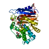
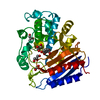
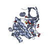
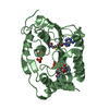
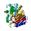
| Title | Toward Better Antibiotics: Crystal Structure Of D-Ala-D-Ala Peptidase inhibited by a novel bicyclic phosphate inhibitor | ||||||
 Components Components | D-alanyl-D-alanine carboxypeptidase | ||||||
 Keywords Keywords | HYDROLASE / CYCLIC PHOSPHATE / PEPTIDOGLYCAN / PENICILLIN BINDING PROTEIN / ANTIBIOTIC | ||||||
| Function / homology |  Function and homology information Function and homology informationserine-type D-Ala-D-Ala carboxypeptidase / serine-type D-Ala-D-Ala carboxypeptidase activity / peptidoglycan biosynthetic process / cell wall organization / regulation of cell shape / proteolysis / extracellular region Similarity search - Function | ||||||
| Biological species |  Streptomyces sp. (bacteria) Streptomyces sp. (bacteria) | ||||||
| Method |  X-RAY DIFFRACTION / SYNCHROTRON / FOURIER SYNTHESIS / Resolution: 1.15 Å X-RAY DIFFRACTION / SYNCHROTRON / FOURIER SYNTHESIS / Resolution: 1.15 Å | ||||||
 Authors Authors | Silvaggi, N.R. / Kaur, K. / Adediran, S.A. / Pratt, R.F. / Kelly, J.A. | ||||||
 Citation Citation | Journal: Biochemistry / Year: 2004 Title: Toward better antibiotics: crystallographic studies of a novel class of DD-peptidase/beta-lactamase inhibitors Authors: Silvaggi, N.R. / Kaur, K. / Adediran, S.A. / Pratt, R.F. / Kelly, J.A. #1: Journal: Biochemistry / Year: 2003Title: Structure of Phosphonate-Inhibited D-Ala-D-Ala Peptidase Reveals an Analogue of a Tetrahedral Transition State. Authors: Silvaggi, N.R. / Anderson, J.W. / Brinsmade, S.R. / Pratt, R.F. / Kelly, J.A. #2: Journal: J.Mol.Biol. / Year: 2002Title: STRUCTURES OF TWO KINETIC INTERMEDIATES REVEAL SPECIES SPECIFICITY OF PENICILLIN-BINDING PROTEINS Authors: McDonough, M.A. / Anderson, J.W. / Silvaggi, N.R. / Pratt, R.F. / Knox, J.R. / Kelly, J.A. #3: Journal: J.Mol.Biol. / Year: 1995Title: THE REFINED CRYSTALLOGRAPHIC STRUCTURE OF A DD-PEPTIDASE PENICILLIN-TARGET ENZYME AT 1.6A RESOLUTION Authors: Kelly, J.A. / Kuzin, A.P. | ||||||
| History |
|
- Structure visualization
| Structure viewer | Molecule: MolmilJmol/JSmol |
|---|
- Downloads & links
-Download
| PDBx/mmCIF format | 1sde.cif.gz | 168.1 KB | Display | PDBx/mmCIF format |
|---|---|---|---|---|
| PDB format | pdb1sde.ent.gz | 131.6 KB | Display | PDB format |
| PDBx/mmJSON format | 1sde.json.gz | Tree view | PDBx/mmJSON format | |
| Others |  Other downloads Other downloads |
-Validation report
| Arichive directory | https://data.pdbj.org/pub/pdb/validation_reports/sd/1sdeftp://data.pdbj.org/pub/pdb/validation_reports/sd/1sde | HTTPS FTP |
|---|
-Related structure data
| Related structure data |  1scwC 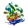 3pteS C: citing same article ( S: Starting model for refinement |
|---|---|
| Similar structure data |


-Links
 PDBj
PDBj
- Assembly
| Deposited unit | 
| ||||||||
|---|---|---|---|---|---|---|---|---|---|
| 1 |
| ||||||||
| Unit cell |
| ||||||||
| Details | Monomer is biologically active unit |
-Components
| #1: Protein | Mass: 37220.371 Da / Num. of mol.: 1 / Source method: isolated from a natural source / Source: (natural) Streptomyces sp. (bacteria) / Strain: R61References: UniProt: P15555, serine-type D-Ala-D-Ala carboxypeptidase |
|---|---|
| #2: Chemical | ChemComp-2PB /   Mass: 200.085 Da / Num. of mol.: 1 / Source method: obtained synthetically / Formula: C7H5O5P Mass: 200.085 Da / Num. of mol.: 1 / Source method: obtained synthetically / Formula: C7H5O5P |
| #3: Chemical | ChemComp-GOL /   Mass: 92.094 Da / Num. of mol.: 1 / Source method: obtained synthetically / Formula: C3H8O3 Mass: 92.094 Da / Num. of mol.: 1 / Source method: obtained synthetically / Formula: C3H8O3 |
| #4: Water | ChemComp-HOH /  Mass: 18.015 Da / Num. of mol.: 568 / Source method: isolated from a natural source / Formula: H2O Mass: 18.015 Da / Num. of mol.: 568 / Source method: isolated from a natural source / Formula: H2O |
| Has protein modification | Y |
-Experimental details
-Experiment
| Experiment | Method: X-RAY DIFFRACTION / Number of used crystals: 1 |
|---|
- Sample preparation
| Crystal | Density Matthews: 2.28 Å3/Da / Density % sol: 46.01 % |
|---|---|
| Crystal grow | Temperature: 298 K / Method: vapor diffusion, hanging drop / pH: 6.8 Details: 20% PEG 8000, 50mM sodium phosphate, pH 6.8, VAPOR DIFFUSION, HANGING DROP, temperature 298.0K |
-Data collection
| Diffraction | Mean temperature: 100 K |
|---|---|
| Diffraction source | Source: SYNCHROTRON / Site: NSLS  / Beamline: X12B / Wavelength: 1 Å / Beamline: X12B / Wavelength: 1 Å |
| Detector | Type: ADSC QUANTUM 315 / Detector: CCD / Date: Jul 14, 2003 / Details: MIRRORS |
| Radiation | Monochromator: SI(111) / Protocol: SINGLE WAVELENGTH / Monochromatic (M) / Laue (L): M / Scattering type: x-ray |
| Radiation wavelength | Wavelength: 1 Å / Relative weight: 1 |
| Reflection | Resolution: 1.15→50 Å / Num. all: 122116 / Num. obs: 119308 / % possible obs: 97.7 % / Observed criterion σ(I): 2 / Redundancy: 6.5 % / Rsym value: 0.056 / Net I/σ(I): 28.8 |
| Reflection shell | Resolution: 1.15→1.19 Å / Redundancy: 4.3 % / Mean I/σ(I) obs: 4.3 / Num. unique all: 9967 / Rsym value: 0.245 / % possible all: 82.9 |
- Processing
| Software |
| |||||||||||||||||||||||||
|---|---|---|---|---|---|---|---|---|---|---|---|---|---|---|---|---|---|---|---|---|---|---|---|---|---|---|
| Refinement | Method to determine structure: FOURIER SYNTHESIS Starting model: 3PTE Resolution: 1.15→10 Å / Num. parameters: 28915 / Num. restraintsaints: 0 / Cross valid method: FREE R / σ(F): 0 / Stereochemistry target values: ENGH AND HUBER Details: ANISOTROPIC REFINEMENT REDUCED FREE R (NO CUTOFF) BY 2.9%
| |||||||||||||||||||||||||
| Displacement parameters | Biso mean: 9.1 Å2 | |||||||||||||||||||||||||
| Refine analyze | Num. disordered residues: 25 / Occupancy sum hydrogen: 0 / Occupancy sum non hydrogen: 3069.67 | |||||||||||||||||||||||||
| Refinement step | Cycle: LAST / Resolution: 1.15→10 Å
| |||||||||||||||||||||||||
| Refine LS restraints |
|
