 Movie
Movie Controller

[English] 日本語
 Yorodumi
- PDB-1pwc: penicilloyl acyl enzyme complex of the Streptomyces R61 DD-peptid... -
+ Open data

- Basic information
| Entry | Database: PDB / ID: 1pwc | ||||||
|---|---|---|---|---|---|---|---|
| Title | penicilloyl acyl enzyme complex of the Streptomyces R61 DD-peptidase with penicillin G | ||||||
 Components Components | D-alanyl-D-alanine carboxypeptidase | ||||||
 Keywords Keywords | HYDROLASE / BETA-LACTAM / ANTIBIOTICS / PENICILLIN BINDING PROTEIN / ENZYME / PEPTIDOGLYCAN | ||||||
| Function / homology |  Function and homology information Function and homology informationserine-type D-Ala-D-Ala carboxypeptidase / serine-type D-Ala-D-Ala carboxypeptidase activity / peptidoglycan biosynthetic process / cell wall organization / regulation of cell shape / proteolysis / extracellular region Similarity search - Function | ||||||
| Biological species |  Streptomyces sp. (bacteria) Streptomyces sp. (bacteria) | ||||||
| Method |  X-RAY DIFFRACTION / SYNCHROTRON / FOURIER SYNTHESIS / Resolution: 1.1 Å X-RAY DIFFRACTION / SYNCHROTRON / FOURIER SYNTHESIS / Resolution: 1.1 Å | ||||||
 Authors Authors | Silvaggi, N.R. / Josephine, H.R. / Pratt, R.F. / Kelly, J.A. | ||||||
 Citation Citation | Journal: J.Mol.Biol. / Year: 2005 Title: Crystal structures of complexes between the R61 DD-peptidase and peptidoglycan-mimetic beta-lactams: a non-covalent complex with a "perfect penicillin" Authors: Silvaggi, N.R. / Josephine, H.R. / Kuzin, A.P. / Nagarajan, R. / Pratt, R.F. / Kelly, J.A. #1: Journal: J.Mol.Biol. / Year: 2002Title: Structures of Two Kinetic Intermediates Reveal Species Specificity of Penicillin-Binding Proteins Authors: Mcdonough, M.A. / Anderson, J.W. / Silvaggi, N.R. / Pratt, R.F. / Knox, J.R. / Kelly, J.A. #2: Journal: Proc.Natl.Acad.Sci.USA / Year: 2001Title: A 1.2-A Snapshot of the Final Step of Bacterial Cell Wall Biosynthesis Authors: Lee, W. / Mcdonough, M.A. / Kotra, L. / Li, Z.H. / Silvaggi, N.R. / Takeda, Y. / Kelly, J.A. / Mobashery, S. #3: Journal: J.Mol.Biol. / Year: 1995Title: The Refined Crystallographic Structure of a Dd-Peptidase Penicillin-Target Enzyme at 1.6 A Resolution Authors: Kelly, J.A. / Kuzin, A.P. | ||||||
| History |
|
- Structure visualization
| Structure viewer | Molecule: MolmilJmol/JSmol |
|---|
- Downloads & links
-Download
| PDBx/mmCIF format | 1pwc.cif.gz | 166.2 KB | Display | PDBx/mmCIF format |
|---|---|---|---|---|
| PDB format | pdb1pwc.ent.gz | 130 KB | Display | PDB format |
| PDBx/mmJSON format | 1pwc.json.gz | Tree view | PDBx/mmJSON format | |
| Others |  Other downloads Other downloads |
-Validation report
| Arichive directory | https://data.pdbj.org/pub/pdb/validation_reports/pw/1pwcftp://data.pdbj.org/pub/pdb/validation_reports/pw/1pwc | HTTPS FTP |
|---|
-Related structure data
| Related structure data | 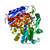 1pw1C  1pw8C 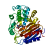 1pwdC 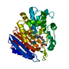 1pwgC 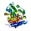 3pteS S: Starting model for refinement C: citing same article ( |
|---|---|
| Similar structure data |


-Links
 PDBj
PDBj
- Assembly
| Deposited unit | 
| ||||||||
|---|---|---|---|---|---|---|---|---|---|
| 1 |
| ||||||||
| Unit cell |
|
-Components
| #1: Protein | Mass: 37422.574 Da / Num. of mol.: 1 / Fragment: DD-PEPTIDASE / Source method: isolated from a natural source / Source: (natural) Streptomyces sp. (bacteria) / Strain: R61References: UniProt: P15555, serine-type D-Ala-D-Ala carboxypeptidase |
|---|---|
| #2: Chemical | ChemComp-PNM /   Mass: 336.406 Da / Num. of mol.: 1 / Source method: obtained synthetically / Formula: C16H20N2O4S Mass: 336.406 Da / Num. of mol.: 1 / Source method: obtained synthetically / Formula: C16H20N2O4S |
| #3: Water | ChemComp-HOH /  Mass: 18.015 Da / Num. of mol.: 507 / Source method: isolated from a natural source / Formula: H2O Mass: 18.015 Da / Num. of mol.: 507 / Source method: isolated from a natural source / Formula: H2O |
| Has protein modification | Y |
-Experimental details
-Experiment
| Experiment | Method: X-RAY DIFFRACTION / Number of used crystals: 1 |
|---|
- Sample preparation
| Crystal | Density Matthews: 1.94 Å3/Da / Density % sol: 45.55 % |
|---|---|
| Crystal grow | Temperature: 298 K / Method: vapor diffusion, hanging drop / pH: 6.8 Details: 20% PEG 8000, 50mM Sodium Phosphate, pH 6.80, VAPOR DIFFUSION, HANGING DROP, temperature 298.0K |
-Data collection
| Diffraction | Mean temperature: 100 K |
|---|---|
| Diffraction source | Source: SYNCHROTRON / Site: NSLS  / Beamline: X12C / Wavelength: 1 Å / Beamline: X12C / Wavelength: 1 Å |
| Detector | Type: BRANDEIS - B4 / Detector: CCD / Date: Apr 14, 2003 / Details: MIRRORS |
| Radiation | Monochromator: SI(111) / Protocol: SINGLE WAVELENGTH / Monochromatic (M) / Laue (L): M / Scattering type: x-ray |
| Radiation wavelength | Wavelength: 1 Å / Relative weight: 1 |
| Reflection | Resolution: 1.1→50 Å / Num. all: 131101 / Num. obs: 131101 / % possible obs: 94.1 % / Observed criterion σ(F): 0 / Observed criterion σ(I): 2 / Redundancy: 4.3 % / Rmerge(I) obs: 0.058 / Net I/σ(I): 14.4 |
| Reflection shell | Resolution: 1.1→1.14 Å / Redundancy: 2.5 % / Rmerge(I) obs: 0.209 / Mean I/σ(I) obs: 3.5 / Num. unique all: 8799 / % possible all: 64.1 |
- Processing
| Software |
| |||||||||||||||||||||||||||||||||
|---|---|---|---|---|---|---|---|---|---|---|---|---|---|---|---|---|---|---|---|---|---|---|---|---|---|---|---|---|---|---|---|---|---|---|
| Refinement | Method to determine structure: FOURIER SYNTHESIS Starting model: 3PTE Resolution: 1.1→10 Å / Num. parameters: 28737 / Num. restraintsaints: 0 / Cross valid method: FREE R / σ(F): 0 / Stereochemistry target values: ENGH AND HUBER Details: ANISOTROPIC REFINEMENT REDUCED FREE R (NO CUTOFF) BY 0.026.
| |||||||||||||||||||||||||||||||||
| Refine analyze | Luzzati coordinate error obs: 0.07 Å / Num. disordered residues: 18 / Occupancy sum hydrogen: 0 / Occupancy sum non hydrogen: 3112.5 | |||||||||||||||||||||||||||||||||
| Refinement step | Cycle: LAST / Resolution: 1.1→10 Å
| |||||||||||||||||||||||||||||||||
| Refine LS restraints |
|
