 Movie
Movie Controller

[English] 日本語
 Yorodumi
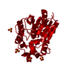
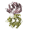
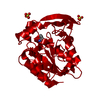
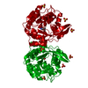
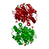
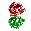
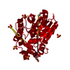
- PDB-1sc9: Hydroxynitrile Lyase from Hevea brasiliensis in complex with the ... -
+ Open data

- Basic information
| Entry | Database: PDB / ID: 1sc9 | ||||||
|---|---|---|---|---|---|---|---|
| Title | Hydroxynitrile Lyase from Hevea brasiliensis in complex with the natural substrate acetone cyanohydrin | ||||||
 Components Components | (S)-acetone-cyanohydrin lyase | ||||||
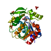
 Keywords Keywords | LYASE / alpha-beta hydrolase fold / substrate complex / catalytic triad | ||||||
| Function / homology |  Function and homology information Function and homology informationaliphatic (S)-hydroxynitrile lyase activity / aromatic (S)-hydroxynitrile lyase activity / (S)-hydroxynitrile lyase / methyl salicylate esterase activity / methyl indole-3-acetate esterase activity / methyl jasmonate esterase activity / salicylic acid metabolic process / jasmonic acid metabolic process Similarity search - Function | ||||||
| Biological species |   Hevea brasiliensis (rubber tree) Hevea brasiliensis (rubber tree) | ||||||
| Method |  X-RAY DIFFRACTION / SYNCHROTRON / FOURIER SYNTHESIS / Resolution: 1.8 Å X-RAY DIFFRACTION / SYNCHROTRON / FOURIER SYNTHESIS / Resolution: 1.8 Å | ||||||
 Authors Authors | Gruber, K. / Gartler, G. / Krammer, B. / Schwab, H. / Kratky, C. | ||||||
 Citation Citation | Journal: J.Biol.Chem. / Year: 2004 Title: Reaction mechanism of hydroxynitrile lyases of the alpha/beta-hydrolase superfamily: the three-dimensional structure of the transient enzyme-substrate complex certifies the crucial role of LYS236 Authors: Gruber, K. / Gartler, G. / Krammer, B. / Schwab, H. / Kratky, C. #1: Journal: Biol.Chem. / Year: 1999Title: Atomic resolution crystal structure of hydroxynitrile lyase from hevea brasiliensis Authors: Gruber, K. / Gugganig, M. / Wagner, U.G. / Kratky, C. #2: Journal: Protein Sci. / Year: 1999Title: Three-dimensional structures of enzyme-substrate complexes of the hydroxynitrile lyase from hevea brasiliensis Authors: Zuegg, J. / Gruber, K. / Gugganig, M. / Wagner, U.G. / Kratky, C. #3: Journal: Structure / Year: 1996Title: Mechanism of cyanogenesis: the crystal structure of hydroxynitrile lyase from Hevea brasiliensis Authors: Wagner, U.G. / Hasslacher, M. / Griengl, H. / Schwab, H. / Kratky, C. | ||||||
| History |
|
- Structure visualization
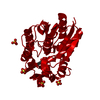
| Structure viewer | Molecule: MolmilJmol/JSmol |
|---|
- Downloads & links
-Download
| PDBx/mmCIF format | 1sc9.cif.gz | 72.9 KB | Display | PDBx/mmCIF format |
|---|---|---|---|---|
| PDB format | pdb1sc9.ent.gz | 53.4 KB | Display | PDB format |
| PDBx/mmJSON format | 1sc9.json.gz | Tree view | PDBx/mmJSON format | |
| Others |  Other downloads Other downloads |
-Validation report
| Arichive directory | https://data.pdbj.org/pub/pdb/validation_reports/sc/1sc9ftp://data.pdbj.org/pub/pdb/validation_reports/sc/1sc9 | HTTPS FTP |
|---|
-Related structure data
| Related structure data |  1sciC 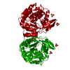 1sckC  1scqC  2yasS S: Starting model for refinement C: citing same article ( |
|---|---|
| Similar structure data |


-Links
 PDBj
PDBj


- Assembly
| Deposited unit | 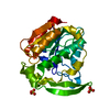
| |||||||||
|---|---|---|---|---|---|---|---|---|---|---|
| 1 | 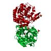
| |||||||||
| 2 |
| |||||||||
| Unit cell |
| |||||||||
| Components on special symmetry positions |
| |||||||||
| Details | dimer; the second part of the biological assembly is generated by the two fod axis: x, -y+1, -z. |
-Components
| #1: Protein | ( Mass: 29262.598 Da / Num. of mol.: 1 Source method: isolated from a genetically manipulated source Source: (gene. exp.) Hevea brasiliensis (rubber tree) / Tissue: leaf / Gene: HNL / Plasmid: BHIL-D2 / Production host:  Pichia pastoris (fungus) / References: UniProt: P52704, EC: 4.1.2.39 Pichia pastoris (fungus) / References: UniProt: P52704, EC: 4.1.2.39 | ||||
|---|---|---|---|---|---|
| #2: Chemical |   Mass: 96.063 Da / Num. of mol.: 2 / Source method: obtained synthetically / Formula: SO4 Mass: 96.063 Da / Num. of mol.: 2 / Source method: obtained synthetically / Formula: SO4#3: Chemical | ChemComp-CNH / | 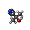  Mass: 85.104 Da / Num. of mol.: 1 / Source method: obtained synthetically / Formula: C4H7NO Mass: 85.104 Da / Num. of mol.: 1 / Source method: obtained synthetically / Formula: C4H7NO#4: Water | ChemComp-HOH / |  Mass: 18.015 Da / Num. of mol.: 310 / Source method: isolated from a natural source / Formula: H2O Mass: 18.015 Da / Num. of mol.: 310 / Source method: isolated from a natural source / Formula: H2O |
-Experimental details
-Experiment
| Experiment | Method: X-RAY DIFFRACTION / Number of used crystals: 1 |
|---|
- Sample preparation
| Crystal | Density Matthews: 2.37 Å3/Da / Density % sol: 47.79 % |
|---|---|
| Crystal grow | Temperature: 298 K / Method: vapor diffusion, hanging drop / pH: 7.5 Details: ammonium sulfate, PEG 400, HEPES, pH 7.5, VAPOR DIFFUSION, HANGING DROP, temperature 298K |
-Data collection
| Diffraction | Mean temperature: 100 K |
|---|---|
| Diffraction source | Source: SYNCHROTRON / Site: EMBL/DESY, HAMBURG  / Beamline: X11 / Wavelength: 0.91 Å / Beamline: X11 / Wavelength: 0.91 Å |
| Detector | Type: MARRESEARCH / Detector: IMAGE PLATE / Date: Jun 8, 2001 |
| Radiation | Monochromator: unknown / Protocol: SINGLE WAVELENGTH / Monochromatic (M) / Laue (L): M / Scattering type: x-ray |
| Radiation wavelength | Wavelength: 0.91 Å / Relative weight: 1 |
| Reflection | Resolution: 1.8→30 Å / Num. all: 30106 / Num. obs: 30106 / % possible obs: 99.1 % / Observed criterion σ(F): 0 / Observed criterion σ(I): 0 / Redundancy: 4.1 % / Biso Wilson estimate: 12.1 Å2 / Rsym value: 0.069 / Net I/σ(I): 19.4 |
| Reflection shell | Resolution: 1.8→1.84 Å / Mean I/σ(I) obs: 6.1 / Num. unique all: 1931 / Rsym value: 0.252 / % possible all: 96.9 |
- Processing
| Software |
| ||||||||||||||||||||||||||||||||||||
|---|---|---|---|---|---|---|---|---|---|---|---|---|---|---|---|---|---|---|---|---|---|---|---|---|---|---|---|---|---|---|---|---|---|---|---|---|---|
| Refinement | Method to determine structure: FOURIER SYNTHESIS Starting model: 2YAS Resolution: 1.8→28.41 Å / Rfactor Rfree error: 0.005 / Data cutoff high absF: 1958997.55 / Data cutoff low absF: 0 / Isotropic thermal model: RESTRAINED / Cross valid method: THROUGHOUT / σ(F): 0 / σ(I): 0 / Stereochemistry target values: Engh & Huber Details: refinement against maximum likelihood target function
| ||||||||||||||||||||||||||||||||||||
| Solvent computation | Solvent model: FLAT MODEL / Bsol: 49.7011 Å2 / ksol: 0.386387 e/Å3 | ||||||||||||||||||||||||||||||||||||
| Displacement parameters | Biso mean: 14.4 Å2
| ||||||||||||||||||||||||||||||||||||
| Refine analyze |
| ||||||||||||||||||||||||||||||||||||
| Refinement step | Cycle: LAST / Resolution: 1.8→28.41 Å
| ||||||||||||||||||||||||||||||||||||
| Refine LS restraints |
| ||||||||||||||||||||||||||||||||||||
| LS refinement shell | Resolution: 1.8→1.84 Å / Rfactor Rfree error: 0.022 / Total num. of bins used: 15
| ||||||||||||||||||||||||||||||||||||
| Xplor file |
|
