 Movie
Movie Controller


+ Open data

- Basic information
| Entry | Database: PDB / ID: 1sac | ||||||
|---|---|---|---|---|---|---|---|
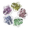
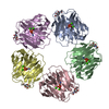
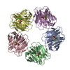
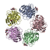
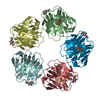
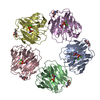
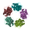
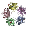
| Title | THE STRUCTURE OF PENTAMERIC HUMAN SERUM AMYLOID P COMPONENT | ||||||
 Components Components | SERUM AMYLOID P COMPONENT | ||||||
 Keywords Keywords | AMYLOID PROTEIN | ||||||
| Function / homology |  Function and homology information Function and homology informationnegative regulation by host of viral glycoprotein metabolic process / negative regulation of glycoprotein metabolic process / complement component C1q complex binding / negative regulation of viral process / negative regulation of wound healing / negative regulation of monocyte differentiation / host-mediated suppression of symbiont invasion / virion binding / negative regulation of acute inflammatory response / chaperone-mediated protein complex assembly ...negative regulation by host of viral glycoprotein metabolic process / negative regulation of glycoprotein metabolic process / complement component C1q complex binding / negative regulation of viral process / negative regulation of wound healing / negative regulation of monocyte differentiation / host-mediated suppression of symbiont invasion / virion binding / negative regulation of acute inflammatory response / chaperone-mediated protein complex assembly / acute-phase response / unfolded protein binding / protein folding / : / carbohydrate binding / blood microparticle / Amyloid fiber formation / innate immune response / calcium ion binding / extracellular space / extracellular exosome / extracellular region / identical protein binding / nucleus Similarity search - Function | ||||||
| Biological species |  Homo sapiens (human) Homo sapiens (human) | ||||||
| Method |  X-RAY DIFFRACTION / Resolution: 2 Å X-RAY DIFFRACTION / Resolution: 2 Å | ||||||
 Authors Authors | White, H.E. / Emsley, J. / O'Hara, B.P. / Oliva, G. / Srinivasan, N. / Tickle, I.J. / Blundell, T.L. / Pepys, M.B. / Wood, S.P. | ||||||
 Citation Citation | Journal: Nature / Year: 1994 Title: Structure of pentameric human serum amyloid P component. Authors: Emsley, J. / White, H.E. / O'Hara, B.P. / Oliva, G. / Srinivasan, N. / Tickle, I.J. / Blundell, T.L. / Pepys, M.B. / Wood, S.P. #1: Journal: J.Mol.Biol. / Year: 1988Title: A Pentameric Form of Human Serum Amyloid P Component: Crystallization, X-Ray Diffraction and Neutron Scattering Studies Authors: Wood, S.P. / Oliva, G. / O'Hara, B.P. / White, H.E. / Blundell, T.L. / Perkins, S.J. / Sardharwalla, I. / Pepys, M.P. #2: Journal: J.Cryst.Growth / Year: 1988Title: Crystallization of Human Serum Amyloid P Component (Sap) Authors: O'Hara, B.P. / Wood, S.P. / Oliva, G. / White, H.E. / Pepys, M.B. #3: Journal: J.Biochem.(Tokyo) / Year: 1986Title: Isolation and Characterization of the Complete Complementary and Genomic DNA Sequences of Human Serum Authors: Ohnishi, S. / Maeda, S. / Shimada, K. / Arao, T. | ||||||
| History |
| ||||||
| Remark 700 | SHEET STRANDS A1_ AND A2_ IN ALL FIVE CHAINS ARE DISCONNECTED BUT FORM A CONTINUOUS STRAND IN THE STRUCTURE. |
- Structure visualization
| Structure viewer | Molecule: MolmilJmol/JSmol |
|---|
- Downloads & links
-Download
| PDBx/mmCIF format | 1sac.cif.gz | 211.2 KB | Display | PDBx/mmCIF format |
|---|---|---|---|---|
| PDB format | pdb1sac.ent.gz | 169.8 KB | Display | PDB format |
| PDBx/mmJSON format | 1sac.json.gz | Tree view | PDBx/mmJSON format | |
| Others |  Other downloads Other downloads |
-Validation report
| Summary document | 1sac_validation.pdf.gz | 411.9 KB | Display | wwPDB validaton report |
|---|---|---|---|---|
| Full document | 1sac_full_validation.pdf.gz | 442.9 KB | Display | |
| Data in XML | 1sac_validation.xml.gz | 24.5 KB | Display | |
| Data in CIF | 1sac_validation.cif.gz | 36 KB | Display | |
| Arichive directory | https://data.pdbj.org/pub/pdb/validation_reports/sa/1sacftp://data.pdbj.org/pub/pdb/validation_reports/sa/1sac | HTTPS FTP |
-Related structure data
| Similar structure data |
|---|

-Links
 PDBj
PDBj

- Assembly
| Deposited unit | 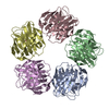
| ||||||||||||||||||||
|---|---|---|---|---|---|---|---|---|---|---|---|---|---|---|---|---|---|---|---|---|---|
| 1 |
| ||||||||||||||||||||
| Unit cell |
| ||||||||||||||||||||
| Atom site foot note | 1: CIS PROLINE - PRO A 89 2: PHE B 33 - THR B 34 OMEGA = 145.17 PEPTIDE BOND DEVIATES SIGNIFICANTLY FROM TRANS CONFORMATION 3: CIS PROLINE - PRO B 89 / 4: CIS PROLINE - PRO C 89 / 5: CIS PROLINE - PRO D 89 / 6: CIS PROLINE - PRO E 89 7: PRO E 129 - LYS E 130 OMEGA = 148.51 PEPTIDE BOND DEVIATES SIGNIFICANTLY FROM TRANS CONFORMATION | ||||||||||||||||||||
| Noncrystallographic symmetry (NCS) | NCS oper:
| ||||||||||||||||||||
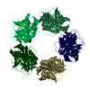
| Details | SAP IS A PENTAMER OF IDENTICAL POLYPEPTIDE CHAINS. THE COORDINATES OF ALL FIVE CHAINS ARE INCLUDED IN THIS ENTRY AND HAVE BEEN ASSIGNED CHAIN INDICATORS *A*, *B*, *C*, *D*, AND *E*. THE TRANSFORMATIONS GIVEN IN THE *MTRIX* RECORDS BELOW REPRESENT THE NON-CRYSTALLOGRAPHIC FIVE-FOLD AXIS. THE TRANSFORMATION PRESENTED ON *MTRIX 1* RECORDS BELOW WILL YIELD APPROXIMATE COORDINATES FOR CHAIN A WHEN APPLIED TO CHAIN B. THE TRANSFORMATION PRESENTED ON *MTRIX 2* RECORDS BELOW WILL YIELD APPROXIMATE COORDINATES FOR CHAIN A WHEN APPLIED TO CHAIN C. THE TRANSFORMATION PRESENTED ON *MTRIX 3* RECORDS BELOW WILL YIELD APPROXIMATE COORDINATES FOR CHAIN A WHEN APPLIED TO CHAIN D. THE TRANSFORMATION PRESENTED ON *MTRIX 4* RECORDS BELOW WILL YIELD APPROXIMATE COORDINATES FOR CHAIN A WHEN APPLIED TO CHAIN E. |
-Components
| #1: Protein | Mass: 23282.455 Da / Num. of mol.: 5 Source method: isolated from a genetically manipulated source Source: (gene. exp.) Homo sapiens (human) / References: UniProt: P02743#2: Chemical | ChemComp-CA /   Mass: 40.078 Da / Num. of mol.: 10 / Source method: obtained synthetically / Formula: Ca Mass: 40.078 Da / Num. of mol.: 10 / Source method: obtained synthetically / Formula: Ca#3: Chemical | ChemComp-ACY /   Mass: 60.052 Da / Num. of mol.: 4 / Source method: obtained synthetically / Formula: C2H4O2 Mass: 60.052 Da / Num. of mol.: 4 / Source method: obtained synthetically / Formula: C2H4O2Has protein modification | Y | Sequence details | SEQUENCE ADVISORY NOTICE DIFFERENCE BETWEEN SWISS-PROT AND PDB SEQUENCE. SWISS-PROT ENTRY NAME: ...SEQUENCE ADVISORY NOTICE DIFFERENCE | |
|---|
-Experimental details
-Experiment
| Experiment | Method: X-RAY DIFFRACTION |
|---|
- Sample preparation
| Crystal | Density Matthews: 2.83 Å3/Da / Density % sol: 56.46 % | ||||||||||||||||||||||||||||||
|---|---|---|---|---|---|---|---|---|---|---|---|---|---|---|---|---|---|---|---|---|---|---|---|---|---|---|---|---|---|---|---|
| Crystal grow | *PLUS Temperature: 4 ℃ / pH: 8 / Method: unknownDetails: taken from Wood, S.P. et al (1988). J. Mol. Biol., 202, 169-173. | ||||||||||||||||||||||||||||||
| Components of the solutions | *PLUS
|
-Data collection
| Reflection | *PLUS Highest resolution: 2 Å / % possible obs: 93.6 % |
|---|
- Processing
| Software |
| ||||||||||||||||||||||||||||||||||||||||||||||||||||||||||||
|---|---|---|---|---|---|---|---|---|---|---|---|---|---|---|---|---|---|---|---|---|---|---|---|---|---|---|---|---|---|---|---|---|---|---|---|---|---|---|---|---|---|---|---|---|---|---|---|---|---|---|---|---|---|---|---|---|---|---|---|---|---|
| Refinement | Resolution: 2→8 Å / σ(F): 0 /
| ||||||||||||||||||||||||||||||||||||||||||||||||||||||||||||
| Refinement step | Cycle: LAST / Resolution: 2→8 Å
| ||||||||||||||||||||||||||||||||||||||||||||||||||||||||||||
| Refine LS restraints |
| ||||||||||||||||||||||||||||||||||||||||||||||||||||||||||||
| Refinement | *PLUS Num. reflection all: 78910 / Rfactor all: 0.179 / Rfactor Rwork: 0.179 | ||||||||||||||||||||||||||||||||||||||||||||||||||||||||||||
| Solvent computation | *PLUS | ||||||||||||||||||||||||||||||||||||||||||||||||||||||||||||
| Displacement parameters | *PLUS | ||||||||||||||||||||||||||||||||||||||||||||||||||||||||||||
| Refine LS restraints | *PLUS Type: x_angle_d / Dev ideal: 3.45 |
