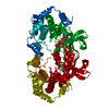
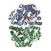
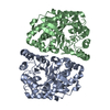
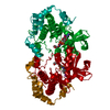
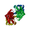
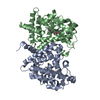
[English] 日本語
Yorodumi
- PDB-1s9i: X-ray structure of the human mitogen-activated protein kinase kin... -

+
Open data


ID or keywords:

Loading...

-
Basic information

Entry
Database: PDB / ID: 1s9i
TitleX-ray structure of the human mitogen-activated protein kinase kinase 2 (MEK2)in a complex with ligand and MgATP
ComponentsDual specificity mitogen-activated protein kinase kinase 2
KeywordsTRANSFERASE / PROTEIN KINASE-LIGAND-MgATP COMPLEX / PROTEIN-PROTEIN INTERACTIONS
Function / homology
Function and homology information


epithelial cell proliferation involved in lung morphogenesis / regulation of axon regeneration / mitogen-activated protein kinase kinase / peptidyl-serine autophosphorylation / MAP-kinase scaffold activity / Signaling by MAP2K mutants / positive regulation of cell motility / regulation of Golgi inheritance / peroxisomal membrane / trachea formation ...epithelial cell proliferation involved in lung morphogenesis / regulation of axon regeneration / mitogen-activated protein kinase kinase / peptidyl-serine autophosphorylation / MAP-kinase scaffold activity / Signaling by MAP2K mutants / positive regulation of cell motility / regulation of Golgi inheritance / peroxisomal membrane / trachea formation / Negative feedback regulation of MAPK pathway / regulation of early endosome to late endosome transport / positive regulation of axonogenesis / regulation of stress-activated MAPK cascade / Frs2-mediated activation / ERBB2-ERBB3 signaling pathway / face development / MAPK1 (ERK2) activation / MAP kinase kinase activity / thyroid gland development / Uptake and function of anthrax toxins / Schwann cell development / ERK1 and ERK2 cascade / protein serine/threonine/tyrosine kinase activity / myelination / protein serine/threonine kinase activator activity / insulin-like growth factor receptor signaling pathway / thymus development / Signal transduction by L1 / PDZ domain binding / RAF activation / Signaling by high-kinase activity BRAF mutants / MAP2K and MAPK activation / positive regulation of protein serine/threonine kinase activity / cytoplasmic side of plasma membrane / Signaling by RAF1 mutants / Signaling by moderate kinase activity BRAF mutants / Paradoxical activation of RAF signaling by kinase inactive BRAF / Signaling downstream of RAS mutants / MAPK cascade / Signaling by BRAF and RAF1 fusions / cell-cell junction / late endosome / heart development / scaffold protein binding / protein tyrosine kinase activity / microtubule / early endosome / protein serine kinase activity / focal adhesion / protein serine/threonine kinase activity / positive regulation of gene expression / positive regulation of DNA-templated transcription / perinuclear region of cytoplasm / Golgi apparatus / endoplasmic reticulum / mitochondrion / extracellular region / ATP binding / nucleus / metal ion binding / cytosol
Similarity search - Function
Transferase(Phosphotransferase) domain 1 / Transferase(Phosphotransferase); domain 1 / Phosphorylase Kinase; domain 1 / Phosphorylase Kinase; domain 1 / Serine/threonine-protein kinase, active site / Serine/Threonine protein kinases active-site signature. / Protein kinase domain / Serine/Threonine protein kinases, catalytic domain / Protein kinase, ATP binding site / Protein kinases ATP-binding region signature. ...Transferase(Phosphotransferase) domain 1 / Transferase(Phosphotransferase); domain 1 / Phosphorylase Kinase; domain 1 / Phosphorylase Kinase; domain 1 / Serine/threonine-protein kinase, active site / Serine/Threonine protein kinases active-site signature. / Protein kinase domain / Serine/Threonine protein kinases, catalytic domain / Protein kinase, ATP binding site / Protein kinases ATP-binding region signature. / Protein kinase domain profile. / Protein kinase domain / Protein kinase-like domain superfamily / 2-Layer Sandwich / Orthogonal Bundle / Mainly Alpha / Alpha Beta
Similarity search - Domain/homology
Chem-5EA / ADENOSINE-5'-TRIPHOSPHATE / Dual specificity mitogen-activated protein kinase kinase 2
Similarity search - Component
Biological speciesHomo sapiens (human)
MethodX-RAY DIFFRACTION / SYNCHROTRON / MOLECULAR REPLACEMENT / Resolution: 3.2 Å
AuthorsOhren, J.F. / Chen, H. / Pavlovsky, A. / Whitehead, C. / Yan, C. / McConnell, P. / Delaney, A. / Dudley, D.T. / Sebolt-Leopold, J. / Hasemann, C.A.
CitationJournal: Nat.Struct.Mol.Biol. / Year: 2004
Title: Structures of human MAP kinase kinase 1 (MEK1) and MEK2 describe novel noncompetitive kinase inhibition.
Authors: Ohren, J.F. / Chen, H. / Pavlovsky, A. / Whitehead, C. / Zhang, E. / Kuffa, P. / Yan, C. / McConnell, P. / Spessard, C. / Banotai, C. / Mueller, W.T. / Delaney, A. / Omer, C. / Sebolt- ...Authors: Ohren, J.F. / Chen, H. / Pavlovsky, A. / Whitehead, C. / Zhang, E. / Kuffa, P. / Yan, C. / McConnell, P. / Spessard, C. / Banotai, C. / Mueller, W.T. / Delaney, A. / Omer, C. / Sebolt-Leopold, J. / Dudley, D.T. / Leung, I.K. / Flamme, C. / Warmus, J. / Kaufman, M. / Barrett, S. / Tecle, H. / Hasemann, C.A.
History
DepositionFeb 4, 2004Deposition site: RCSB / Processing site: RCSB
Revision 1.0Nov 23, 2004Provider: repository / Type: Initial release
Revision 1.1Apr 29, 2008Group: Version format compliance
Revision 1.2Jul 13, 2011Group: Version format compliance
Revision 1.3Feb 14, 2024Group: Data collection / Database references / Derived calculations
Category: chem_comp_atom / chem_comp_bond ...chem_comp_atom / chem_comp_bond / database_2 / pdbx_struct_conn_angle / struct_conn / struct_ref_seq_dif / struct_site
Item: _database_2.pdbx_DOI / _database_2.pdbx_database_accession ..._database_2.pdbx_DOI / _database_2.pdbx_database_accession / _pdbx_struct_conn_angle.ptnr1_auth_comp_id / _pdbx_struct_conn_angle.ptnr1_auth_seq_id / _pdbx_struct_conn_angle.ptnr1_label_asym_id / _pdbx_struct_conn_angle.ptnr1_label_atom_id / _pdbx_struct_conn_angle.ptnr1_label_comp_id / _pdbx_struct_conn_angle.ptnr1_label_seq_id / _pdbx_struct_conn_angle.ptnr3_auth_comp_id / _pdbx_struct_conn_angle.ptnr3_auth_seq_id / _pdbx_struct_conn_angle.ptnr3_label_asym_id / _pdbx_struct_conn_angle.ptnr3_label_atom_id / _pdbx_struct_conn_angle.ptnr3_label_comp_id / _pdbx_struct_conn_angle.ptnr3_label_seq_id / _pdbx_struct_conn_angle.value / _struct_conn.pdbx_dist_value / _struct_conn.ptnr1_auth_comp_id / _struct_conn.ptnr1_auth_seq_id / _struct_conn.ptnr1_label_asym_id / _struct_conn.ptnr1_label_atom_id / _struct_conn.ptnr1_label_comp_id / _struct_conn.ptnr1_label_seq_id / _struct_conn.ptnr2_auth_comp_id / _struct_conn.ptnr2_auth_seq_id / _struct_conn.ptnr2_label_asym_id / _struct_conn.ptnr2_label_atom_id / _struct_conn.ptnr2_label_comp_id / _struct_conn.ptnr2_label_seq_id / _struct_ref_seq_dif.details / _struct_site.pdbx_auth_asym_id / _struct_site.pdbx_auth_comp_id / _struct_site.pdbx_auth_seq_id
Revision 1.4Apr 3, 2024Group: Refinement description / Category: pdbx_initial_refinement_model

-
Structure visualization

Structure viewerMolecule:
MolmilJmol/JSmol

Downloads & links

-
Assembly

Deposited unit
A: Dual specificity mitogen-activated protein kinase kinase 2
B: Dual specificity mitogen-activated protein kinase kinase 2
hetero molecules


Theoretical massNumber of molelcules
Total (without water)81,3468
Polymers79,1932
Non-polymers2,1546
Water00
1


  • Idetical with deposited unit
  • defined by author&software
TypeNameSymmetry operationNumber
identity operation1_555x,y,z1
Buried area5500 Å2
ΔGint-31 kcal/mol
Surface area28410 Å2
MethodPISA
Unit cell
Length a, b, c (Å)161.888, 161.888, 122.986
Angle α, β, γ (deg.)90.00, 90.00, 120.00
Int Tables number178
Space group name H-MP6122
DetailsThe NH2-truncated MEK2 is a homodimer in solution and in the crystallographic asu. MEK2 may form a functional dimer in vivo.

-
Components

#1: Protein Dual specificity mitogen-activated protein kinase kinase 2 / MAP kinase kinase 2 / MAPKK 2 / ERK activator kinase 2 / MAPK/ERK kinase 2 / MEK2


Mass: 39596.363 Da / Num. of mol.: 2
Source method: isolated from a genetically manipulated source
Source: (gene. exp.) Homo sapiens (human) / Gene: MAP2K2, PRKMK2, MEK2, MKK2 / Plasmid: pET24b / Species (production host): Escherichia coli / Production host: Escherichia coli BL21(DE3) (bacteria) / Strain (production host): BL21(DE3) / References: UniProt: P36507, EC: 2.7.1.37
#2: Chemical ChemComp-MG / MAGNESIUM ION


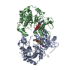
Mass: 24.305 Da / Num. of mol.: 2 / Source method: obtained synthetically / Formula: Mg
#3: Chemical ChemComp-ATP / ADENOSINE-5'-TRIPHOSPHATE


Mass: 507.181 Da / Num. of mol.: 2 / Source method: obtained synthetically / Formula: C10H16N5O13P3 / Comment: ATP, energy-carrying molecule*YM
#4: Chemical ChemComp-5EA / 5-{3,4-DIFLUORO-2-[(2-FLUORO-4-IODOPHENYL)AMINO]PHENYL}-N-(2-MORPHOLIN-4-YLETHYL)-1,3,4-OXADIAZOL-2-AMINE


Mass: 545.297 Da / Num. of mol.: 2 / Source method: obtained synthetically / Formula: C20H19F3IN5O2

-
Experimental details

-
Experiment

ExperimentMethod: X-RAY DIFFRACTION / Number of used crystals: 1

-
Sample preparation

CrystalDensity Matthews: 2.94 Å3/Da / Density % sol: 58.12 %
Crystal growTemperature: 288 K / pH: 7
Details: Sodium / Potassium phosphate, DTT, pH 7, VAPOR DIFFUSION, HANGING DROP, temperature 288K, pH 7.00

-
Data collection

DiffractionMean temperature: 100 K
Diffraction sourceSource: SYNCHROTRON / Site: APS / Beamline: 17-ID / Wavelength: 1
DetectorType: MARRESEARCH / Detector: CCD / Date: Aug 2, 2002
RadiationMonochromator: SAGITALLY FOCUSED SI(111) / Protocol: SINGLE WAVELENGTH / Monochromatic (M) / Laue (L): M / Scattering type: x-ray
Radiation wavelengthWavelength: 1 Å / Relative weight: 1
ReflectionResolution: 3.2→14.4 Å / Num. obs: 15147 / % possible obs: 99.9 % / Observed criterion σ(I): 0 / Redundancy: 9.8 % / Rmerge(I) obs: 0.088 / Net I/σ(I): 25.6
Reflection shellResolution: 3.2→3.28 Å / Rmerge(I) obs: 0.368 / Mean I/σ(I) obs: 6 / % possible all: 1

-
Processing

Software
NameVersionClassification
HKL-2000data collection
TRUNCATEdata reduction
MOLREPphasing
REFMAC5.1.24refinement
HKL-2000data reduction
CCP4(TRUNCATE)data scaling
RefinementMethod to determine structure: MOLECULAR REPLACEMENT
Starting model: HUMAN MAP2K1

Resolution: 3.2→14.4 Å / Cor.coef. Fo:Fc: 0.85 / Cor.coef. Fo:Fc free: 0.772 / SU B: 30.11 / SU ML: 0.534 / Isotropic thermal model: Isotropic / Cross valid method: THROUGHOUT / ESU R Free: 0.667 / Stereochemistry target values: MAXIMUM LIKELIHOOD / Details: MAXIMUM LIKELIHOOD REFINEMENT USING REFMAC 5.1.24
RfactorNum. reflection% reflectionSelection details
Rfree0.36374 802 5 %RANDOM
Rwork0.29028 ---
obs0.29401 15147 99.91 %-
Solvent computationIon probe radii: 0.8 Å / Shrinkage radii: 0.8 Å / VDW probe radii: 1.4 Å / Solvent model: BABINET MODEL WITH MASK
Displacement parametersBiso mean: 41.694 Å2
Baniso -1Baniso -2Baniso -3
1--1.36 Å2-0.68 Å20 Å2
2---1.36 Å20 Å2
3---2.04 Å2
Refinement stepCycle: LAST / Resolution: 3.2→14.4 Å
ProteinNucleic acidLigandSolventTotal
Num. atoms4686 0 126 0 4812
Refine LS restraints
Refine-IDTypeDev idealDev ideal targetNumber
X-RAY DIFFRACTIONr_bond_refined_d0.0070.0214889
X-RAY DIFFRACTIONr_bond_other_d
X-RAY DIFFRACTIONr_angle_refined_deg1.2171.9996601
X-RAY DIFFRACTIONr_angle_other_deg
X-RAY DIFFRACTIONr_dihedral_angle_1_deg5.4355588
X-RAY DIFFRACTIONr_dihedral_angle_2_deg
X-RAY DIFFRACTIONr_chiral_restr0.0720.2718
X-RAY DIFFRACTIONr_gen_planes_refined0.0020.023655
X-RAY DIFFRACTIONr_gen_planes_other
X-RAY DIFFRACTIONr_nbd_refined0.1950.22388
X-RAY DIFFRACTIONr_nbd_other
X-RAY DIFFRACTIONr_nbtor_other
X-RAY DIFFRACTIONr_xyhbond_nbd_refined0.130.2142
X-RAY DIFFRACTIONr_xyhbond_nbd_other
X-RAY DIFFRACTIONr_symmetry_vdw_refined0.1820.230
X-RAY DIFFRACTIONr_symmetry_vdw_other
X-RAY DIFFRACTIONr_symmetry_hbond_refined0.0490.21
X-RAY DIFFRACTIONr_symmetry_hbond_other
X-RAY DIFFRACTIONr_mcbond_it0.3551.52953
X-RAY DIFFRACTIONr_mcangle_it0.65924774
X-RAY DIFFRACTIONr_scbond_it0.64631936
X-RAY DIFFRACTIONr_scangle_it1.1754.51827
X-RAY DIFFRACTIONr_rigid_bond_restr
X-RAY DIFFRACTIONr_sphericity_free
X-RAY DIFFRACTIONr_sphericity_bonded
LS refinement shellResolution: 3.201→3.28 Å / Total num. of bins used: 20 /
RfactorNum. reflection
Rfree0.482 52
Rwork0.335 1055

+
About Yorodumi

-
News

-
Feb 9, 2022. New format data for meta-information of EMDB entries

New format data for meta-information of EMDB entries

  • Version 3 of the EMDB header file is now the official format.
  • The previous official version 1.9 will be removed from the archive.

Related info.:EMDB header

External links:wwPDB to switch to version 3 of the EMDB data model

-
Aug 12, 2020. Covid-19 info

Covid-19 info

URL: https://pdbjlvh1.pdbj.org/emnavi/covid19.php

New page: Covid-19 featured information page in EM Navigator.

Related info.:Covid-19 info / Mar 5, 2020. Novel coronavirus structure data

+
Mar 5, 2020. Novel coronavirus structure data

Novel coronavirus structure data

Related info.:Yorodumi Speices / Aug 12, 2020. Covid-19 info

External links:COVID-19 featured content - PDBj / Molecule of the Month (242):Coronavirus Proteases

+
Jan 31, 2019. EMDB accession codes are about to change! (news from PDBe EMDB page)

EMDB accession codes are about to change! (news from PDBe EMDB page)

  • The allocation of 4 digits for EMDB accession codes will soon come to an end. Whilst these codes will remain in use, new EMDB accession codes will include an additional digit and will expand incrementally as the available range of codes is exhausted. The current 4-digit format prefixed with “EMD-” (i.e. EMD-XXXX) will advance to a 5-digit format (i.e. EMD-XXXXX), and so on. It is currently estimated that the 4-digit codes will be depleted around Spring 2019, at which point the 5-digit format will come into force.
  • The EM Navigator/Yorodumi systems omit the EMD- prefix.

Related info.:Q: What is EMD? / ID/Accession-code notation in Yorodumi/EM Navigator

External links:EMDB Accession Codes are Changing Soon! / Contact to PDBj

+
Jul 12, 2017. Major update of PDB

Major update of PDB

  • wwPDB released updated PDB data conforming to the new PDBx/mmCIF dictionary.
  • This is a major update changing the version number from 4 to 5, and with Remediation, in which all the entries are updated.
  • In this update, many items about electron microscopy experimental information are reorganized (e.g. em_software).
  • Now, EM Navigator and Yorodumi are based on the updated data.

External links:wwPDB Remediation / Enriched Model Files Conforming to OneDep Data Standards Now Available in the PDB FTP Archive

-
Yorodumi

Thousand views of thousand structures

  • Yorodumi is a browser for structure data from EMDB, PDB, SASBDB, etc.
  • This page is also the successor to EM Navigator detail page, and also detail information page/front-end page for Omokage search.
  • The word "yorodu" (or yorozu) is an old Japanese word meaning "ten thousand". "mi" (miru) is to see.

Related info.:EMDB / PDB / SASBDB / Comparison of 3 databanks / Yorodumi Search / Aug 31, 2016. New EM Navigator & Yorodumi / Yorodumi Papers / Jmol/JSmol / Function and homology information / Changes in new EM Navigator and Yorodumi

Read more