 Movie
Movie Controller

[English] 日本語
 Yorodumi
- PDB-1s6m: Conjugative Relaxase Trwc In Complex With Orit DNA. Metal-Bound S... -
+ Open data

- Basic information
| Entry | Database: PDB / ID: 1s6m | ||||||
|---|---|---|---|---|---|---|---|
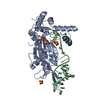
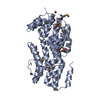
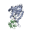
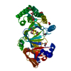
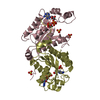
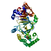
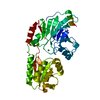
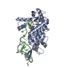
| Title | Conjugative Relaxase Trwc In Complex With Orit DNA. Metal-Bound Structure | ||||||
 Components Components |
| ||||||
 Keywords Keywords | DNA BINDING PROTEIN/DNA / protein-dna complex / bacterial conjugation / relaxase / dna replication / DNA BINDING PROTEIN-DNA COMPLEX | ||||||
| Function / homology |  Function and homology information Function and homology information | ||||||
| Biological species |  | ||||||
| Method |  X-RAY DIFFRACTION / SYNCHROTRON / MOLECULAR REPLACEMENT / Resolution: 2.28 Å X-RAY DIFFRACTION / SYNCHROTRON / MOLECULAR REPLACEMENT / Resolution: 2.28 Å | ||||||
 Authors Authors | Guasch, A. / Lucas, M. / Moncalian, G. / Cabezas, M. / Perez-Luque, R. / Gomis-Ruth, F.X. / de la Cruz, F. / Coll, M. | ||||||
 Citation Citation | Journal: J.Mol.Biol. / Year: 2006 Title: Unveiling the molecular mechanism of a conjugative relaxase: The structure of TrwC complexed with a 27-mer DNA comprising the recognition hairpin and the cleavage site. Authors: Boer, R. / Russi, S. / Guasch, A. / Lucas, M. / Blanco, A.G. / Perez-Luque, R. / Coll, M. / de la Cruz, F. | ||||||
| History |
|
- Structure visualization
| Structure viewer | Molecule: MolmilJmol/JSmol |
|---|
- Downloads & links
-Download
| PDBx/mmCIF format | 1s6m.cif.gz | 88.8 KB | Display | PDBx/mmCIF format |
|---|---|---|---|---|
| PDB format | pdb1s6m.ent.gz | 64.8 KB | Display | PDB format |
| PDBx/mmJSON format | 1s6m.json.gz | Tree view | PDBx/mmJSON format | |
| Others |  Other downloads Other downloads |
-Validation report
| Arichive directory | https://data.pdbj.org/pub/pdb/validation_reports/s6/1s6mftp://data.pdbj.org/pub/pdb/validation_reports/s6/1s6m | HTTPS FTP |
|---|
-Related structure data
| Related structure data |  1zm5C  2cdmC  1omhS S: Starting model for refinement C: citing same article ( |
|---|---|
| Similar structure data |


-Links
 PDBj
PDBj


- Assembly
| Deposited unit | 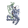
| ||||||||
|---|---|---|---|---|---|---|---|---|---|
| 1 |
| ||||||||
| Unit cell |
|
-Components
| #1: DNA chain | Mass: 7714.970 Da / Num. of mol.: 1 / Source method: obtained synthetically |
|---|---|
| #2: Protein | Mass: 33082.414 Da / Num. of mol.: 1 Source method: isolated from a genetically manipulated source Source: (gene. exp.) |
| #3: Chemical | ChemComp-NI /   Mass: 58.693 Da / Num. of mol.: 1 / Source method: obtained synthetically / Formula: Ni Mass: 58.693 Da / Num. of mol.: 1 / Source method: obtained synthetically / Formula: Ni |
| #4: Water | ChemComp-HOH /  Mass: 18.015 Da / Num. of mol.: 162 / Source method: isolated from a natural source / Formula: H2O Mass: 18.015 Da / Num. of mol.: 162 / Source method: isolated from a natural source / Formula: H2O |
| Has protein modification | Y |
-Experimental details
-Experiment
| Experiment | Method: X-RAY DIFFRACTION / Number of used crystals: 1 |
|---|
- Sample preparation
| Crystal | Density Matthews: 3.18 Å3/Da / Density % sol: 61.28 % |
|---|---|
| Crystal grow | Temperature: 296 K / pH: 4.6 Details: 25% PEGMM 2000, 0.2 M AMMONIUM SULPHATE, 0.1 M SODIUM ACETATE, PH 4.6, VAPOR DIFFUSION, HANGING DROP, TEMPERATURE 296K Temp details: K |
-Data collection
| Diffraction | Mean temperature: 110 K |
|---|---|
| Diffraction source | Source: SYNCHROTRON / Site: ESRF  / Beamline: BM14 / Wavelength: 0.983994 Å / Beamline: BM14 / Wavelength: 0.983994 Å |
| Detector | Type: MARRESEARCH / Detector: CCD / Date: Mar 19, 2003 |
| Radiation | Protocol: SINGLE WAVELENGTH / Monochromatic (M) / Laue (L): M / Scattering type: x-ray |
| Radiation wavelength | Wavelength: 0.983994 Å / Relative weight: 1 |
| Reflection | Resolution: 2.28→45.264 Å / Num. all: 24644 / Num. obs: 24598 / % possible obs: 100 % / Observed criterion σ(F): 1 / Observed criterion σ(I): 1 |
- Processing
| Software |
| ||||||||||||||||||||
|---|---|---|---|---|---|---|---|---|---|---|---|---|---|---|---|---|---|---|---|---|---|
| Refinement | Method to determine structure: MOLECULAR REPLACEMENT Starting model: 1omh Resolution: 2.28→43.85 Å / Cross valid method: THROUGHOUT / σ(F): 0 / Stereochemistry target values: Engh & Huber
| ||||||||||||||||||||
| Refinement step | Cycle: LAST / Resolution: 2.28→43.85 Å
| ||||||||||||||||||||
| Refine LS restraints |
|
