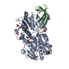
 Movie
Movie Controller

[English] 日本語
 Yorodumi
- PDB-1m7h: Crystal Structure of APS kinase from Penicillium Chrysogenum: Str... -
+ Open data

- Basic information
| Entry | Database: PDB / ID: 1m7h | ||||||
|---|---|---|---|---|---|---|---|
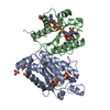
| Title | Crystal Structure of APS kinase from Penicillium Chrysogenum: Structure with APS soaked out of one dimer | ||||||
 Components Components | Adenylylsulfate kinase | ||||||
 Keywords Keywords | TRANSFERASE / APS kinase / Adenylylsulfate kinase / sulfate metabolism / Nucleotide 2 kinase | ||||||
| Function / homology |  Function and homology information Function and homology informationadenylyl-sulfate kinase / adenylylsulfate kinase activity / sulfate assimilation / hydrogen sulfide biosynthetic process / L-cysteine biosynthetic process / : / ATP binding / cytosol Similarity search - Function | ||||||
| Biological species |  Penicillium chrysogenum (fungus) Penicillium chrysogenum (fungus) | ||||||
| Method |  X-RAY DIFFRACTION / SYNCHROTRON / MOLECULAR REPLACEMENT / Resolution: 2 Å X-RAY DIFFRACTION / SYNCHROTRON / MOLECULAR REPLACEMENT / Resolution: 2 Å | ||||||
 Authors Authors | Lansdon, E.B. / Sege, I.H. / Fisher, A.J. | ||||||
 Citation Citation | Journal: Biochemistry / Year: 2002 Title: Ligand-Induced Structural Changes in Adenosine 5'-Phosphosulfate Kinase from Penicillium chrysogenum. Authors: Lansdon, E.B. / Segel, I.H. / Fisher, A.J. | ||||||
| History |
| ||||||
| Remark 999 | SEQUENCE Residue 54 in all of the subunits is listed as Arg in the database sequence (SwissProt ...SEQUENCE Residue 54 in all of the subunits is listed as Arg in the database sequence (SwissProt accession Q12657). According to the author, the gene has been resequenced and residue 54 is actually Gly. |
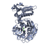
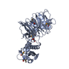
- Structure visualization
| Structure viewer | Molecule: MolmilJmol/JSmol |
|---|
- Downloads & links
-Download
| PDBx/mmCIF format | 1m7h.cif.gz | 185.3 KB | Display | PDBx/mmCIF format |
|---|---|---|---|---|
| PDB format | pdb1m7h.ent.gz | 148.4 KB | Display | PDB format |
| PDBx/mmJSON format | 1m7h.json.gz | Tree view | PDBx/mmJSON format | |
| Others |  Other downloads Other downloads |
-Validation report
| Arichive directory | https://data.pdbj.org/pub/pdb/validation_reports/m7/1m7hftp://data.pdbj.org/pub/pdb/validation_reports/m7/1m7h | HTTPS FTP |
|---|
-Related structure data


-Links
 PDBj
PDBj
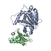
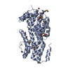
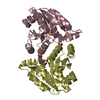
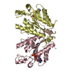
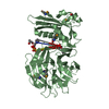
- Assembly
| Deposited unit | 
| ||||||||
|---|---|---|---|---|---|---|---|---|---|
| 1 | 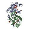
| ||||||||
| 2 | 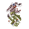
| ||||||||
| 3 | 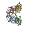
| ||||||||
| 4 | 
| ||||||||
| Unit cell |
|
-Components
| #1: Protein | Mass: 23804.781 Da / Num. of mol.: 4 Source method: isolated from a genetically manipulated source Source: (gene. exp.) Penicillium chrysogenum (fungus) / Plasmid: pET23A / Species (production host): Escherichia coli / Production host:  #2: Chemical | ChemComp-SO4 /   Mass: 96.063 Da / Num. of mol.: 12 / Source method: obtained synthetically / Formula: SO4 Mass: 96.063 Da / Num. of mol.: 12 / Source method: obtained synthetically / Formula: SO4#3: Chemical | ChemComp-ADP /   Mass: 427.201 Da / Num. of mol.: 4 / Source method: obtained synthetically / Formula: C10H15N5O10P2 / Comment: ADP, energy-carrying molecule*YM Mass: 427.201 Da / Num. of mol.: 4 / Source method: obtained synthetically / Formula: C10H15N5O10P2 / Comment: ADP, energy-carrying molecule*YM#4: Chemical |   Type: RNA linking / Mass: 427.284 Da / Num. of mol.: 2 / Source method: obtained synthetically / Formula: C10H14N5O10PS Type: RNA linking / Mass: 427.284 Da / Num. of mol.: 2 / Source method: obtained synthetically / Formula: C10H14N5O10PS#5: Water | ChemComp-HOH / |  Mass: 18.015 Da / Num. of mol.: 499 / Source method: isolated from a natural source / Formula: H2O Mass: 18.015 Da / Num. of mol.: 499 / Source method: isolated from a natural source / Formula: H2O |
|---|
-Experimental details
-Experiment
| Experiment | Method: X-RAY DIFFRACTION / Number of used crystals: 1 |
|---|
- Sample preparation
| Crystal | Density Matthews: 2.55 Å3/Da / Density % sol: 51.69 % | ||||||||||||||||||||||||||||||||||||||||||
|---|---|---|---|---|---|---|---|---|---|---|---|---|---|---|---|---|---|---|---|---|---|---|---|---|---|---|---|---|---|---|---|---|---|---|---|---|---|---|---|---|---|---|---|
| Crystal grow | Temperature: 298 K / Method: vapor diffusion, hanging drop / pH: 4 Details: Na monobasic phosphate, K dibasic phosphate, succinate, pH 4, VAPOR DIFFUSION, HANGING DROP, temperature 298K | ||||||||||||||||||||||||||||||||||||||||||
| Crystal grow | *PLUS pH: 8 | ||||||||||||||||||||||||||||||||||||||||||
| Components of the solutions | *PLUS
|
-Data collection
| Diffraction | Mean temperature: 100 K |
|---|---|
| Diffraction source | Source: SYNCHROTRON / Site: SSRL  / Beamline: BL9-2 / Wavelength: 0.98 Å / Beamline: BL9-2 / Wavelength: 0.98 Å |
| Detector | Type: ADSC QUANTUM 4 / Detector: CCD / Date: Jan 29, 2002 |
| Radiation | Monochromator: double crystal / Protocol: SINGLE WAVELENGTH / Monochromatic (M) / Laue (L): M / Scattering type: x-ray |
| Radiation wavelength | Wavelength: 0.98 Å / Relative weight: 1 |
| Reflection | Resolution: 2→30 Å / Num. all: 621412 / Num. obs: 621412 / % possible obs: 97.3 % / Observed criterion σ(I): -3 / Redundancy: 9.61 % / Biso Wilson estimate: 20.4 Å2 / Rmerge(I) obs: 0.05 / Rsym value: 0.05 / Net I/σ(I): 14.1 |
| Reflection shell | Resolution: 2→2.05 Å / Rmerge(I) obs: 0.329 / Mean I/σ(I) obs: 3.7 / Num. unique all: 4004 / Rsym value: 0.329 / % possible all: 91.5 |
| Reflection | *PLUS Lowest resolution: 30 Å / Num. obs: 64678 / Num. measured all: 621412 / Rmerge(I) obs: 0.05 |
| Reflection shell | *PLUS % possible obs: 91.5 % |
- Processing
| Software |
| |||||||||||||||||||||||||
|---|---|---|---|---|---|---|---|---|---|---|---|---|---|---|---|---|---|---|---|---|---|---|---|---|---|---|
| Refinement | Method to determine structure: MOLECULAR REPLACEMENT Starting model: Ternary structure of APS kinase Resolution: 2→29.91 Å / Rfactor Rfree error: 0.005 / Isotropic thermal model: RESTRAINED / Cross valid method: THROUGHOUT / σ(F): 0 / Stereochemistry target values: Engh & Huber
| |||||||||||||||||||||||||
| Solvent computation | Solvent model: FLAT MODEL / Bsol: 54.4752 Å2 / ksol: 0.369587 e/Å3 | |||||||||||||||||||||||||
| Displacement parameters | Biso mean: 38.4 Å2
| |||||||||||||||||||||||||
| Refine analyze | Luzzati coordinate error free: 0.31 Å / Luzzati sigma a free: 0.25 Å | |||||||||||||||||||||||||
| Refinement step | Cycle: LAST / Resolution: 2→29.91 Å
| |||||||||||||||||||||||||
| Refine LS restraints |
| |||||||||||||||||||||||||
| LS refinement shell | Resolution: 2→2.13 Å / Rfactor Rfree error: 0.014 / Total num. of bins used: 6
| |||||||||||||||||||||||||
| Xplor file |
| |||||||||||||||||||||||||
| Refinement | *PLUS Lowest resolution: 30 Å / Num. reflection obs: 64678 / % reflection Rfree: 5 % | |||||||||||||||||||||||||
| Solvent computation | *PLUS | |||||||||||||||||||||||||
| Displacement parameters | *PLUS | |||||||||||||||||||||||||
| Refine LS restraints | *PLUS
|
