 Movie
Movie Controller

[English] 日本語
 Yorodumi
- PDB-1s31: Crystal Structure Analysis of the human Tub protein (isoform a) s... -
+ Open data

- Basic information
| Entry | Database: PDB / ID: 1s31 | ||||||
|---|---|---|---|---|---|---|---|
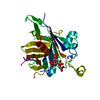
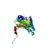
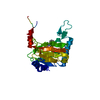
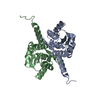
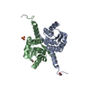
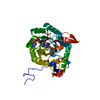
| Title | Crystal Structure Analysis of the human Tub protein (isoform a) spanning residues 289 through 561 | ||||||
 Components Components | tubby isoform a | ||||||
 Keywords Keywords | SIGNALING PROTEIN / BETA BARREL / HYDROPHOBIC HELIX / HYDROPHOBIC CORE | ||||||
| Function / homology |  Function and homology information Function and homology informationintraciliary transport particle A binding / protein localization to photoreceptor outer segment / receptor localization to non-motile cilium / intraciliary transport / phagocytosis, recognition / protein localization to cilium / regulation of G protein-coupled receptor signaling pathway / photoreceptor cell maintenance / positive regulation of phagocytosis / sensory perception of sound ...intraciliary transport particle A binding / protein localization to photoreceptor outer segment / receptor localization to non-motile cilium / intraciliary transport / phagocytosis, recognition / protein localization to cilium / regulation of G protein-coupled receptor signaling pathway / photoreceptor cell maintenance / positive regulation of phagocytosis / sensory perception of sound / G protein-coupled receptor binding / retina development in camera-type eye / cilium / protein-containing complex binding / extracellular region / nucleus / plasma membrane / cytoplasm / cytosol Similarity search - Function | ||||||
| Biological species |  Homo sapiens (human) Homo sapiens (human) | ||||||
| Method |  X-RAY DIFFRACTION / SYNCHROTRON / MOLECULAR REPLACEMENT / molecular replacement / Resolution: 2.704 Å X-RAY DIFFRACTION / SYNCHROTRON / MOLECULAR REPLACEMENT / molecular replacement / Resolution: 2.704 Å | ||||||
 Authors Authors | Boutboul, S. / Carroll, K.J. / Basdevant, A. / Gomez, C. / Nandrot, E. / Clement, K. / Shapiro, L. / Abitbol, M. | ||||||
 Citation Citation | Journal: To be Published Title: A novel human obesity and sensory deficit syndrome resulting from a mutation in the TUB gene Authors: Boutboul, S. / Carroll, K.J. / Basdevant, A. / Gomez, C. / Nandrot, E. / Clement, K. / Shapiro, L. / Abitbol, M. #1: Journal: METHODS ENZYMOL. / Year: 1997Title: Processing of X-ray Diffraction Data Collected in Oscillation Mode Authors: Otwinowski, Z. / Minor, W. #2: Journal: ACTA CRYSTALLOGR.,SECT.A / Year: 1994Title: AMoRe: an Automated Package for Molecular Replacement Authors: Navaza, J. #3: Journal: ACTA CRYSTALLOGR.,SECT.D / Year: 1997Title: Refinement of Macromolecular Structures by the Maximum-Likelihood Method Authors: Murshudov, G.N. / Vagin, A.A. / Dodson, E.J. | ||||||
| History |
|
- Structure visualization
| Structure viewer | Molecule: MolmilJmol/JSmol |
|---|
- Downloads & links
-Download
| PDBx/mmCIF format | 1s31.cif.gz | 71.2 KB | Display | PDBx/mmCIF format |
|---|---|---|---|---|
| PDB format | pdb1s31.ent.gz | 51.4 KB | Display | PDB format |
| PDBx/mmJSON format | 1s31.json.gz | Tree view | PDBx/mmJSON format | |
| Others |  Other downloads Other downloads |
-Validation report
| Arichive directory | https://data.pdbj.org/pub/pdb/validation_reports/s3/1s31ftp://data.pdbj.org/pub/pdb/validation_reports/s3/1s31 | HTTPS FTP |
|---|
-Related structure data
| Related structure data | 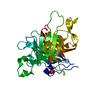 1c8zS S: Starting model for refinement |
|---|---|
| Similar structure data |


-Links
 PDBj
PDBj- Assembly
| Deposited unit | 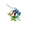
| ||||||||
|---|---|---|---|---|---|---|---|---|---|
| 1 |
| ||||||||
| 2 | 
| ||||||||
| Unit cell |
|
-Components
| #1: Protein | Mass: 30798.025 Da / Num. of mol.: 1 Source method: isolated from a genetically manipulated source Source: (gene. exp.) Homo sapiens (human) / Gene: tub / Plasmid: pSMT3 / Species (production host): Escherichia coli / Production host:  |
|---|---|
| #2: Chemical | ChemComp-PGE /   Mass: 150.173 Da / Num. of mol.: 1 / Source method: obtained synthetically / Formula: C6H14O4 Mass: 150.173 Da / Num. of mol.: 1 / Source method: obtained synthetically / Formula: C6H14O4 |
| #3: Water | ChemComp-HOH /  Mass: 18.015 Da / Num. of mol.: 90 / Source method: isolated from a natural source / Formula: H2O Mass: 18.015 Da / Num. of mol.: 90 / Source method: isolated from a natural source / Formula: H2O |
-Experimental details
-Experiment
| Experiment | Method: X-RAY DIFFRACTION / Number of used crystals: 1 |
|---|
- Sample preparation
| Crystal | Density Matthews: 4.56 Å3/Da / Density % sol: 73.05 % |
|---|---|
| Crystal grow | Temperature: 277.2 K / Method: vapor diffusion, hanging drop / pH: 5 Details: 16% PEG 400, 5mM Magnesium Chloride, 100mM Sodium Acetate, 15mM DTT, VAPOR DIFFUSION, HANGING DROP, temperature 277.2K, pH 5.0 |
-Data collection
| Diffraction | Mean temperature: 110 K |
|---|---|
| Diffraction source | Source: SYNCHROTRON / Site: APS  / Beamline: 32-ID / Wavelength: 0.9814 Å / Beamline: 32-ID / Wavelength: 0.9814 Å |
| Detector | Type: MARRESEARCH / Detector: CCD / Date: Aug 23, 2003 |
| Radiation | Monochromator: Diamond / Protocol: SINGLE WAVELENGTH / Monochromatic (M) / Laue (L): M / Scattering type: x-ray |
| Radiation wavelength | Wavelength: 0.9814 Å / Relative weight: 1 |
| Reflection | Resolution: 2.7→20 Å / Num. all: 15560 / Num. obs: 15503 / % possible obs: 100 % / Observed criterion σ(F): 0 / Observed criterion σ(I): -3 / Redundancy: 24 % / Rmerge(I) obs: 0.096 / Net I/σ(I): 30.9 |
| Reflection shell | Resolution: 2.7→2.8 Å / % possible obs: 100 % / Rmerge(I) obs: 0.344 / Mean I/σ(I) obs: 10.1 / Num. unique all: 1511 / % possible all: 99.7 |
-Phasing
| Phasing | Method: molecular replacement | |||||||||
|---|---|---|---|---|---|---|---|---|---|---|
| Phasing MR | Rfactor: 31.7 / Cor.coef. Fo:Fc: 72.5
|
- Processing
| Software |
| |||||||||||||||||||||||||||||||||||||||||||||||||||||||||||||||||||||||||||||||||||||||||||||||||||||||||||||||||||||
|---|---|---|---|---|---|---|---|---|---|---|---|---|---|---|---|---|---|---|---|---|---|---|---|---|---|---|---|---|---|---|---|---|---|---|---|---|---|---|---|---|---|---|---|---|---|---|---|---|---|---|---|---|---|---|---|---|---|---|---|---|---|---|---|---|---|---|---|---|---|---|---|---|---|---|---|---|---|---|---|---|---|---|---|---|---|---|---|---|---|---|---|---|---|---|---|---|---|---|---|---|---|---|---|---|---|---|---|---|---|---|---|---|---|---|---|---|---|---|
| Refinement | Method to determine structure: MOLECULAR REPLACEMENT Starting model: PDB ENTRY 1C8Z Resolution: 2.704→18.474 Å / Cor.coef. Fo:Fc: 0.946 / Cor.coef. Fo:Fc free: 0.918 / SU B: 14.137 / SU ML: 0.155 / SU R Cruickshank DPI: 0.312 / TLS residual ADP flag: LIKELY RESIDUAL / Cross valid method: THROUGHOUT / σ(F): 0 / σ(I): 0 / ESU R Free: 0.243 / Stereochemistry target values: Engh & Huber / Details: HYDROGENS HAVE BEEN ADDED IN THE RIDING POSITIONS
| |||||||||||||||||||||||||||||||||||||||||||||||||||||||||||||||||||||||||||||||||||||||||||||||||||||||||||||||||||||
| Solvent computation | Ion probe radii: 0.8 Å / Shrinkage radii: 0.8 Å / VDW probe radii: 1.2 Å / Solvent model: BABINET MODEL PLUS MASK | |||||||||||||||||||||||||||||||||||||||||||||||||||||||||||||||||||||||||||||||||||||||||||||||||||||||||||||||||||||
| Displacement parameters | Biso mean: 33.802 Å2 | |||||||||||||||||||||||||||||||||||||||||||||||||||||||||||||||||||||||||||||||||||||||||||||||||||||||||||||||||||||
| Refinement step | Cycle: LAST / Resolution: 2.704→18.474 Å
| |||||||||||||||||||||||||||||||||||||||||||||||||||||||||||||||||||||||||||||||||||||||||||||||||||||||||||||||||||||
| Refine LS restraints |
|