intraciliary transport particle A binding / protein localization to photoreceptor outer segment / receptor localization to non-motile cilium / intraciliary transport / phagocytosis, recognition / protein localization to cilium / regulation of G protein-coupled receptor signaling pathway / photoreceptor cell maintenance / positive regulation of phagocytosis / sensory perception of sound ...intraciliary transport particle A binding / protein localization to photoreceptor outer segment / receptor localization to non-motile cilium / intraciliary transport / phagocytosis, recognition / protein localization to cilium / regulation of G protein-coupled receptor signaling pathway / photoreceptor cell maintenance / positive regulation of phagocytosis / sensory perception of sound / G protein-coupled receptor binding / retina development in camera-type eye / cilium / protein-containing complex binding / extracellular region / nucleus / plasma membrane / cytoplasm / cytosol Similarity search - Function
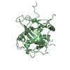
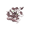
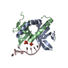
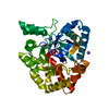
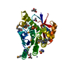
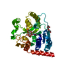
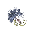
Tubby Protein; Chain A / Tubby Protein; Chain A / Tubby N-terminal / Tubby, C-terminal / Tubby, C-terminal, conserved site / Tub family / Tub family signature 1. / Tub family signature 2. / Tubby, N-terminal / Tubby-like, C-terminal ...Tubby Protein; Chain A / Tubby Protein; Chain A / Tubby N-terminal / Tubby, C-terminal / Tubby, C-terminal, conserved site / Tub family / Tub family signature 1. / Tub family signature 2. / Tubby, N-terminal / Tubby-like, C-terminal / Alpha-Beta Barrel / Alpha Beta Similarity search - Domain/homology
In the structure databanks used in Yorodumi, some data are registered as the other names, "COVID-19 virus" and "2019-nCoV". Here are the details of the virus and the list of structure data.
Jan 31, 2019. EMDB accession codes are about to change! (news from PDBe EMDB page)
EMDB accession codes are about to change! (news from PDBe EMDB page)
The allocation of 4 digits for EMDB accession codes will soon come to an end. Whilst these codes will remain in use, new EMDB accession codes will include an additional digit and will expand incrementally as the available range of codes is exhausted. The current 4-digit format prefixed with “EMD-” (i.e. EMD-XXXX) will advance to a 5-digit format (i.e. EMD-XXXXX), and so on. It is currently estimated that the 4-digit codes will be depleted around Spring 2019, at which point the 5-digit format will come into force.
The EM Navigator/Yorodumi systems omit the EMD- prefix.
Related info.:Q: What is EMD? / ID/Accession-code notation in Yorodumi/EM Navigator
Yorodumi is a browser for structure data from EMDB, PDB, SASBDB, etc.
This page is also the successor to EM Navigator detail page, and also detail information page/front-end page for Omokage search.
The word "yorodu" (or yorozu) is an old Japanese word meaning "ten thousand". "mi" (miru) is to see.
Related info.:EMDB / PDB / SASBDB / Comparison of 3 databanks / Yorodumi Search / Aug 31, 2016. New EM Navigator & Yorodumi / Yorodumi Papers / Jmol/JSmol / Function and homology information / Changes in new EM Navigator and Yorodumi
 Movie
Movie Controller


 Open data
 Basic information
 Components
 Keywords
 Function and homology information
 X-RAY DIFFRACTION /
  Authors
 Citation
 Structure visualization
 Downloads & links
 Other downloads
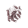

 PDBj
PDBj Assembly
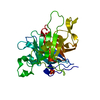


 Mass: 94.971 Da / Num. of mol.: 2 / Source method: obtained synthetically / Formula: PO4
 Mass: 18.015 Da / Num. of mol.: 509 / Source method: isolated from a natural source / Formula: H2O
 Sample preparation
 / Beamline: X4A / Wavelength: 0.9537
  Processing