 Movie
Movie Controller


+ Open data

- Basic information
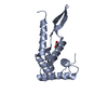
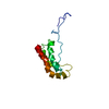
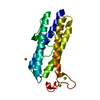
| Entry | Database: PDB / ID: 1rss | ||||||
|---|---|---|---|---|---|---|---|
| Title | RIBOSOMAL PROTEIN S7 FROM THERMUS THERMOPHILUS | ||||||
 Components Components | RIBOSOMAL PROTEIN S7 | ||||||
 Keywords Keywords | RIBOSOMAL PROTEIN / RNA-BINDING / TRANSLATION | ||||||
| Function / homology |  Function and homology information Function and homology informationsmall ribosomal subunit / tRNA binding / rRNA binding / structural constituent of ribosome / translation Similarity search - Function | ||||||
| Biological species |   Thermus thermophilus (bacteria) Thermus thermophilus (bacteria) | ||||||
| Method |  X-RAY DIFFRACTION / SYNCHROTRON / MAD / Resolution: 1.9 Å X-RAY DIFFRACTION / SYNCHROTRON / MAD / Resolution: 1.9 Å | ||||||
 Authors Authors | Wimberly, B. / White, S. / Ramakrishnan, V. | ||||||
 Citation Citation | Journal: Structure / Year: 1997 Title: The structure of ribosomal protein S7 at 1.9 A resolution reveals a beta-hairpin motif that binds double-stranded nucleic acids. Authors: Wimberly, B.T. / White, S.W. / Ramakrishnan, V. | ||||||
| History |
|
- Structure visualization
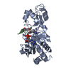
| Structure viewer | Molecule: MolmilJmol/JSmol |
|---|
- Downloads & links
-Download
| PDBx/mmCIF format | 1rss.cif.gz | 42.9 KB | Display | PDBx/mmCIF format |
|---|---|---|---|---|
| PDB format | pdb1rss.ent.gz | 29.9 KB | Display | PDB format |
| PDBx/mmJSON format | 1rss.json.gz | Tree view | PDBx/mmJSON format | |
| Others |  Other downloads Other downloads |
-Validation report
| Arichive directory | https://data.pdbj.org/pub/pdb/validation_reports/rs/1rssftp://data.pdbj.org/pub/pdb/validation_reports/rs/1rss | HTTPS FTP |
|---|
-Related structure data
| Similar structure data |
|---|


-Links
 PDBj
PDBj


- Assembly
| Deposited unit | 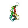
| ||||||||
|---|---|---|---|---|---|---|---|---|---|
| 1 |
| ||||||||
| Unit cell |
|
-Components
| #1: Protein | Mass: 17351.115 Da / Num. of mol.: 1 / Mutation: 5 RESIDUES DELETED FROM N-TERMINUS Source method: isolated from a genetically manipulated source Source: (gene. exp.) Thermus thermophilus (bacteria) / Cell line: BL21 / Gene: S7 / Plasmid: PET-13A / Species (production host): Escherichia coli / Gene (production host): S7 / Production host: |
|---|---|
| #2: Water | ChemComp-HOH /  Mass: 18.015 Da / Num. of mol.: 146 / Source method: isolated from a natural source / Formula: H2O Mass: 18.015 Da / Num. of mol.: 146 / Source method: isolated from a natural source / Formula: H2O |
-Experimental details
-Experiment
| Experiment | Method: X-RAY DIFFRACTION / Number of used crystals: 1 |
|---|
- Sample preparation
| Crystal | Density Matthews: 2.7 Å3/Da / Density % sol: 40 % / Description: 2 WAVELENGTH MAD EXPERIMENT | ||||||||||||||||||||||||||||||||||||||||||||||||||||||
|---|---|---|---|---|---|---|---|---|---|---|---|---|---|---|---|---|---|---|---|---|---|---|---|---|---|---|---|---|---|---|---|---|---|---|---|---|---|---|---|---|---|---|---|---|---|---|---|---|---|---|---|---|---|---|---|
| Crystal grow | Temperature: 277 K / pH: 8 Details: CRYSTALLIZED FROM 15% PEG 8000, 20% GLYCEROL, 6% MPD, 0.1 M NAHCO3, 0.1 M TRIS PH 8.0 AT 4 C, temperature 277K | ||||||||||||||||||||||||||||||||||||||||||||||||||||||
| Crystal grow | *PLUS Temperature: 4-13 ℃ / pH: 7.5 / Method: vapor diffusion, hanging drop | ||||||||||||||||||||||||||||||||||||||||||||||||||||||
| Components of the solutions | *PLUS
|
-Data collection
| Diffraction | Mean temperature: 100 K | |||||||||
|---|---|---|---|---|---|---|---|---|---|---|
| Diffraction source | Source: SYNCHROTRON / Site: NSLS  / Beamline: X12C / Wavelength: 0.98 / Wavelength: 0.98, 1.01 / Beamline: X12C / Wavelength: 0.98 / Wavelength: 0.98, 1.01 | |||||||||
| Detector | Type: BRANDEIS / Detector: CCD / Date: May 15, 1997 | |||||||||
| Radiation | Monochromatic (M) / Laue (L): M / Scattering type: x-ray | |||||||||
| Radiation wavelength |
| |||||||||
| Reflection | Resolution: 1.9→100 Å / Num. obs: 13428 / % possible obs: 99.8 % / Observed criterion σ(I): 0 / Redundancy: 7.7 % / Biso Wilson estimate: 25.9 Å2 / Rmerge(I) obs: 0.033 / Rsym value: 0.04 / Net I/σ(I): 37 | |||||||||
| Reflection shell | Resolution: 1.9→1.98 Å / Redundancy: 5 % / Rmerge(I) obs: 0.064 / Mean I/σ(I) obs: 5 / Rsym value: 0.307 / % possible all: 95 | |||||||||
| Reflection | *PLUS Num. measured all: 103923 |
- Processing
| Software |
| ||||||||||||||||||||||||||||||||||||||||||||||||||||||||||||||||||||||||||||||||
|---|---|---|---|---|---|---|---|---|---|---|---|---|---|---|---|---|---|---|---|---|---|---|---|---|---|---|---|---|---|---|---|---|---|---|---|---|---|---|---|---|---|---|---|---|---|---|---|---|---|---|---|---|---|---|---|---|---|---|---|---|---|---|---|---|---|---|---|---|---|---|---|---|---|---|---|---|---|---|---|---|---|
| Refinement | Method to determine structure: MAD / Resolution: 1.9→6 Å / Rfactor Rfree error: 0.008 / Data cutoff high absF: 1000000 / Data cutoff low absF: 0.001 / Isotropic thermal model: RESTRAINED / Cross valid method: THROUGHOUT / σ(F): 0
| ||||||||||||||||||||||||||||||||||||||||||||||||||||||||||||||||||||||||||||||||
| Displacement parameters | Biso mean: 35.9 Å2
| ||||||||||||||||||||||||||||||||||||||||||||||||||||||||||||||||||||||||||||||||
| Refine analyze |
| ||||||||||||||||||||||||||||||||||||||||||||||||||||||||||||||||||||||||||||||||
| Refinement step | Cycle: LAST / Resolution: 1.9→6 Å
| ||||||||||||||||||||||||||||||||||||||||||||||||||||||||||||||||||||||||||||||||
| Refine LS restraints |
| ||||||||||||||||||||||||||||||||||||||||||||||||||||||||||||||||||||||||||||||||
| LS refinement shell | Resolution: 1.9→2.01 Å / Rfactor Rfree error: 0.022 / Total num. of bins used: 6
| ||||||||||||||||||||||||||||||||||||||||||||||||||||||||||||||||||||||||||||||||
| Xplor file |
| ||||||||||||||||||||||||||||||||||||||||||||||||||||||||||||||||||||||||||||||||
| Software | *PLUS Name: X-PLOR / Version: 3.843 / Classification: refinement | ||||||||||||||||||||||||||||||||||||||||||||||||||||||||||||||||||||||||||||||||
| Refine LS restraints | *PLUS
|
