 Movie
Movie Controller


+ Open data

- Basic information
| Entry | Database: PDB / ID: 1rq2 | ||||||
|---|---|---|---|---|---|---|---|
| Title | MYCOBACTERIUM TUBERCULOSIS FTSZ IN COMPLEX WITH CITRATE | ||||||
 Components Components | Cell division protein ftsZ | ||||||
 Keywords Keywords | CELL CYCLE / SIGNALING PROTEIN / TUBULIN / GTPASE | ||||||
| Function / homology |  Function and homology information Function and homology informationseptin ring assembly / division septum assembly / FtsZ-dependent cytokinesis / cell division site / protein polymerization / positive regulation of cell cycle / cell division / GTPase activity / GTP binding / magnesium ion binding ...septin ring assembly / division septum assembly / FtsZ-dependent cytokinesis / cell division site / protein polymerization / positive regulation of cell cycle / cell division / GTPase activity / GTP binding / magnesium ion binding / plasma membrane / cytoplasm Similarity search - Function | ||||||
| Biological species |   Mycobacterium tuberculosis (bacteria) Mycobacterium tuberculosis (bacteria) | ||||||
| Method |  X-RAY DIFFRACTION / SYNCHROTRON / MOLECULAR REPLACEMENT / Resolution: 1.86 Å X-RAY DIFFRACTION / SYNCHROTRON / MOLECULAR REPLACEMENT / Resolution: 1.86 Å | ||||||
 Authors Authors | Leung, A.K.W. / White, E.L. / Ross, L.J. / Reynolds, R.C. / DeVito, J.A. / Borhani, D.W. | ||||||
 Citation Citation | Journal: J.Mol.Biol. / Year: 2004 Title: Structure of Mycobacterium tuberculosis FtsZ reveals unexpected, G protein-like conformational switches. Authors: Leung, A.K. / Lucile White, E. / Ross, L.J. / Reynolds, R.C. / DeVito, J.A. / Borhani, D.W. #1: Journal: To be PublishedTitle: Polymerization of C-Terminally Truncated Mycobacterium tuberculosis FtsZ Is Unlikely to be Physiologically Relevant Authors: Borhani, D.W. / White, E.L. #2: Journal: Acta Crystallogr.,Sect.D / Year: 2000 Title: Crystallization of the Mycobacterium tuberculosis cell-division protein FtsZ Authors: Leung, A.K.W. / White, E.L. / Ross, L.J. / Borhani, D.W. #3: Journal: J.Bacteriol. / Year: 2000 Title: Slow polymerization of Mycobacterium tuberculosis FtsZ Authors: White, E.L. / Ross, L.J. / Reynolds, R.C. / Seitz, L.E. / Moore, G.D. / Borhani, D.W. #4: Journal: J.ANTIMICROB.CHEMOTHER. / Year: 2002 Title: 2-Alkoxycarbonylaminopyridines: inhibitors of Mycobacterium tuberculosis FtsZ Authors: White, E.L. / Suling, W.J. / Ross, L.J. / Seitz, L.E. / Reynolds, R.C. | ||||||
| History |
|
- Structure visualization
| Structure viewer | Molecule: MolmilJmol/JSmol |
|---|
- Downloads & links
-Download
| PDBx/mmCIF format | 1rq2.cif.gz | 128.3 KB | Display | PDBx/mmCIF format |
|---|---|---|---|---|
| PDB format | pdb1rq2.ent.gz | 98.5 KB | Display | PDB format |
| PDBx/mmJSON format | 1rq2.json.gz | Tree view | PDBx/mmJSON format | |
| Others |  Other downloads Other downloads |
-Validation report
| Arichive directory | https://data.pdbj.org/pub/pdb/validation_reports/rq/1rq2ftp://data.pdbj.org/pub/pdb/validation_reports/rq/1rq2 | HTTPS FTP |
|---|
-Related structure data
| Related structure data | 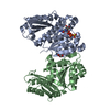 1rluC  1rq7C  1fszS C: citing same article ( S: Starting model for refinement |
|---|---|
| Similar structure data |


-Links
 PDBj
PDBj


- Assembly
| Deposited unit | 
| ||||||||
|---|---|---|---|---|---|---|---|---|---|
| 1 |
| ||||||||
| Unit cell |
|
-Components
| #1: Protein | Mass: 39073.004 Da / Num. of mol.: 2 Source method: isolated from a genetically manipulated source Source: (gene. exp.) Mycobacterium tuberculosis (bacteria) / Gene: FTSZ, RV2150C, MT2209, MTCY270.18, MB2174C / Plasmid: pJD168 / Production host: #2: Chemical | ChemComp-CIT / |   Mass: 192.124 Da / Num. of mol.: 1 / Source method: obtained synthetically / Formula: C6H8O7 Mass: 192.124 Da / Num. of mol.: 1 / Source method: obtained synthetically / Formula: C6H8O7#3: Water | ChemComp-HOH / |  Mass: 18.015 Da / Num. of mol.: 311 / Source method: isolated from a natural source / Formula: H2O Mass: 18.015 Da / Num. of mol.: 311 / Source method: isolated from a natural source / Formula: H2O |
|---|
-Experimental details
-Experiment
| Experiment | Method: X-RAY DIFFRACTION / Number of used crystals: 1 |
|---|
- Sample preparation
| Crystal | Density Matthews: 2.58 Å3/Da / Density % sol: 53 % |
|---|---|
| Crystal grow | Temperature: 293 K / pH: 5.6 Details: 30% PEG 4000, 0.1M SODIUM CITRATE, 0.2M AMMONIUM ACETATE, 2MM SRI-7614, ETHYL (6-AMINO-2,3-DIHYDRO-4-PHENYL-1H-PYRIDO[4,3-B][1,4]DIAZEPIN-8-YL)-CARBAMATE, WAS INCLUDED AS WELL, BUT WAS NOT ...Details: 30% PEG 4000, 0.1M SODIUM CITRATE, 0.2M AMMONIUM ACETATE, 2MM SRI-7614, ETHYL (6-AMINO-2,3-DIHYDRO-4-PHENYL-1H-PYRIDO[4,3-B][1,4]DIAZEPIN-8-YL)-CARBAMATE, WAS INCLUDED AS WELL, BUT WAS NOT LOCATED IN THE FINAL STRUCTURE, VAPOR DIFFUSION, SITTING DROP, temperature 293K, pH 5.60 |
-Data collection
| Diffraction | Mean temperature: 100 K |
|---|---|
| Diffraction source | Source: SYNCHROTRON / Site: CHESS  / Beamline: A1 / Wavelength: 0.9235 / Beamline: A1 / Wavelength: 0.9235 |
| Detector | Type: ADSC QUANTUM 1 / Detector: CCD / Date: Jul 30, 2000 / Details: MIRRORS/MONOCHROMATOR |
| Radiation | Monochromator: SI 111 / Protocol: SINGLE WAVELENGTH / Monochromatic (M) / Laue (L): M / Scattering type: x-ray |
| Radiation wavelength | Wavelength: 0.9235 Å / Relative weight: 1 |
| Reflection | Resolution: 1.86→30 Å / Num. obs: 68271 / Observed criterion σ(I): 0 / Redundancy: 3.5 % / Biso Wilson estimate: 25.68 Å2 / Rmerge(I) obs: 0.061 / Rsym value: 0.061 / Net I/σ(I): 11.4 |
| Reflection shell | Resolution: 1.86→1.93 Å / Redundancy: 3.1 % / Rmerge(I) obs: 0.409 / Mean I/σ(I) obs: 1.3 / Rsym value: 0.409 / % possible all: 99.1 |
- Processing
| Software |
| ||||||||||||||||||||||||||||||||||||||||||||||||||||||||||||||||||||||||||||||||||||||||||||||||||||||||||||||||||||||||||||||||||||||||||||||||||||||||||||||||
|---|---|---|---|---|---|---|---|---|---|---|---|---|---|---|---|---|---|---|---|---|---|---|---|---|---|---|---|---|---|---|---|---|---|---|---|---|---|---|---|---|---|---|---|---|---|---|---|---|---|---|---|---|---|---|---|---|---|---|---|---|---|---|---|---|---|---|---|---|---|---|---|---|---|---|---|---|---|---|---|---|---|---|---|---|---|---|---|---|---|---|---|---|---|---|---|---|---|---|---|---|---|---|---|---|---|---|---|---|---|---|---|---|---|---|---|---|---|---|---|---|---|---|---|---|---|---|---|---|---|---|---|---|---|---|---|---|---|---|---|---|---|---|---|---|---|---|---|---|---|---|---|---|---|---|---|---|---|---|---|---|---|
| Refinement | Method to determine structure: MOLECULAR REPLACEMENT Starting model: PDB ENTRY 1FSZ Resolution: 1.86→30 Å / Cor.coef. Fo:Fc: 0.956 / Cor.coef. Fo:Fc free: 0.944 / SU B: 2.621 / SU ML: 0.079 / Isotropic thermal model: Isotropic / Cross valid method: THROUGHOUT / ESU R: 0.116 / ESU R Free: 0.115 / Stereochemistry target values: MAXIMUM LIKELIHOOD Details: X-PLOR-GENERATED BULK SOLVENT PARTIAL STRUCTURE FACTORS WERE USED IN REFMAC AS PARTIAL STRUCTURE FACTORS (FPART/PHIPART), EXCEPT IN LAST ROUND, WHERE REFMAC BABINET MODEL WITH MASK WAS USED. ...Details: X-PLOR-GENERATED BULK SOLVENT PARTIAL STRUCTURE FACTORS WERE USED IN REFMAC AS PARTIAL STRUCTURE FACTORS (FPART/PHIPART), EXCEPT IN LAST ROUND, WHERE REFMAC BABINET MODEL WITH MASK WAS USED. AFTER COMPLETION OF THE REFINEMENT, AN UNINTERPRETABLE STRETCH OF ELECTRON DENSITY REMAINED NEAR THE ACTIVE SITE OF SUBUNIT A. THIS DENSITY IS DISTINCT FROM THE CITRATE (AND SURROUNDING WATER MOLECULES) IN THE ACTIVE SITE. NONE OF THE OTHER CRYSTALLIZATION BUFFER COMPONENTS COULD BE MODELED INTO THIS DENSITY. A THREE OR FOUR RESIDUE PEPTIDE COULD BE MODELED INTO THIS DENSITY, BUT NOT IN A VERY SATISFACTORY MANNER. THE DENSITY MAY ARISE FROM PARTIAL ORDERING OF THE AMINO-TERMINUS OF ANOTHER FTSZ MOLECULE IN THE CRYSTAL LATTICE, OR MORE LIKELY FROM ONE OF THE PEPTIDIC PROTEASE INHIBITORS INCLUDED IN THE PROTEIN PREPARATION (E.G., LEUPEPTIN).
| ||||||||||||||||||||||||||||||||||||||||||||||||||||||||||||||||||||||||||||||||||||||||||||||||||||||||||||||||||||||||||||||||||||||||||||||||||||||||||||||||
| Solvent computation | Ion probe radii: 0.8 Å / Shrinkage radii: 0.8 Å / VDW probe radii: 1.4 Å / Solvent model: BABINET MODEL WITH MASK | ||||||||||||||||||||||||||||||||||||||||||||||||||||||||||||||||||||||||||||||||||||||||||||||||||||||||||||||||||||||||||||||||||||||||||||||||||||||||||||||||
| Displacement parameters | Biso mean: 28.217 Å2
| ||||||||||||||||||||||||||||||||||||||||||||||||||||||||||||||||||||||||||||||||||||||||||||||||||||||||||||||||||||||||||||||||||||||||||||||||||||||||||||||||
| Refinement step | Cycle: LAST / Resolution: 1.86→30 Å
| ||||||||||||||||||||||||||||||||||||||||||||||||||||||||||||||||||||||||||||||||||||||||||||||||||||||||||||||||||||||||||||||||||||||||||||||||||||||||||||||||
| Refine LS restraints |
| ||||||||||||||||||||||||||||||||||||||||||||||||||||||||||||||||||||||||||||||||||||||||||||||||||||||||||||||||||||||||||||||||||||||||||||||||||||||||||||||||
| LS refinement shell | Resolution: 1.86→1.908 Å / Total num. of bins used: 20 /
|
