 Movie
Movie Controller

[English] 日本語
 Yorodumi
- PDB-1qz0: Crystal Structure of the Yersinia Pestis Phosphatase YopH in Comp... -
+ Open data

- Basic information
| Entry | Database: PDB / ID: 1qz0 | ||||||
|---|---|---|---|---|---|---|---|
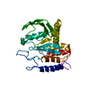
| Title | Crystal Structure of the Yersinia Pestis Phosphatase YopH in Complex with a Phosphotyrosyl Mimetic-Containing Hexapeptide | ||||||
 Components Components |
| ||||||
 Keywords Keywords | HYDROLASE/HYDROLASE INHIBITOR / PHOSPHATASE / PTPASE / YOPH / DEPHOSPHORYLASE / HYDROLASE-HYDROLASE INHIBITOR COMPLEX | ||||||
| Function / homology |  Function and homology information Function and homology informationsymbiont-mediated suppression of host reactive oxygen species generation / symbiont-mediated disruption of host focal adhesion / protein-tyrosine-phosphatase / protein tyrosine phosphatase activity / extracellular region Similarity search - Function | ||||||
| Biological species |   Yersinia pestis (bacteria) Yersinia pestis (bacteria) | ||||||
| Method |  X-RAY DIFFRACTION / SYNCHROTRON / MOLECULAR REPLACEMENT / Resolution: 1.5 Å X-RAY DIFFRACTION / SYNCHROTRON / MOLECULAR REPLACEMENT / Resolution: 1.5 Å | ||||||
 Authors Authors | Phan, J. / Lee, K. / Cherry, S. / Tropea, J.E. / Burke Jr, T.R. / Waugh, D.S. | ||||||
 Citation Citation | Journal: Biochemistry / Year: 2003 Title: High-Resolution Structure of the Yersinia pestis Protein Tyrosine Phosphatase YopH in Complex with a Phosphotyrosyl Mimetic-Containing Hexapeptide Authors: Phan, J. / Lee, K. / Cherry, S. / Tropea, J.E. / Burke Jr, T.R. / Waugh, D.S. | ||||||
| History |
|
- Structure visualization
| Structure viewer | Molecule: MolmilJmol/JSmol |
|---|
- Downloads & links
-Download
| PDBx/mmCIF format | 1qz0.cif.gz | 138.4 KB | Display | PDBx/mmCIF format |
|---|---|---|---|---|
| PDB format | pdb1qz0.ent.gz | 106.5 KB | Display | PDB format |
| PDBx/mmJSON format | 1qz0.json.gz | Tree view | PDBx/mmJSON format | |
| Others |  Other downloads Other downloads |
-Validation report
| Arichive directory | https://data.pdbj.org/pub/pdb/validation_reports/qz/1qz0ftp://data.pdbj.org/pub/pdb/validation_reports/qz/1qz0 | HTTPS FTP |
|---|
-Related structure data
| Related structure data |  1yptS S: Starting model for refinement |
|---|---|
| Similar structure data |


-Links
 PDBj
PDBj


- Assembly
| Deposited unit | 
| ||||||||
|---|---|---|---|---|---|---|---|---|---|
| 1 | 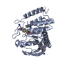
| ||||||||
| 2 | 
| ||||||||
| Unit cell |
|
-Components
| #1: Protein | Mass: 33553.883 Da / Num. of mol.: 2 / Fragment: Catalytic Domain, Residues 164-468 / Mutation: C235R Source method: isolated from a genetically manipulated source Source: (gene. exp.) Yersinia pestis (bacteria) / Plasmid: pZZ1089 / Species (production host): Escherichia coli / Production host: References: GenBank: 16082755, UniProt: O68720*PLUS, protein-tyrosine-phosphatase #2: Protein/peptide | 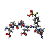  Type: Peptide-like / Class: Inhibitor / Mass: 836.710 Da / Num. of mol.: 4 / Source method: obtained synthetically / References: Ac-DADE-F(2)Pmp-L-NH(2) Type: Peptide-like / Class: Inhibitor / Mass: 836.710 Da / Num. of mol.: 4 / Source method: obtained synthetically / References: Ac-DADE-F(2)Pmp-L-NH(2)#3: Water | ChemComp-HOH / |  Mass: 18.015 Da / Num. of mol.: 603 / Source method: isolated from a natural source / Formula: H2O Mass: 18.015 Da / Num. of mol.: 603 / Source method: isolated from a natural source / Formula: H2OHas protein modification | Y | |
|---|
-Experimental details
-Experiment
| Experiment | Method: X-RAY DIFFRACTION / Number of used crystals: 1 |
|---|
- Sample preparation
| Crystal | Density Matthews: 2.25 Å3/Da / Density % sol: 45.25 % | ||||||||||||||||||||||||||||||
|---|---|---|---|---|---|---|---|---|---|---|---|---|---|---|---|---|---|---|---|---|---|---|---|---|---|---|---|---|---|---|---|
| Crystal grow | Temperature: 291 K / Method: vapor diffusion, hanging drop / pH: 7.5 Details: 20% PEG 3350, 100 mM HEPES, 100 mM NaCl, pH 7.5, VAPOR DIFFUSION, HANGING DROP, temperature 291K | ||||||||||||||||||||||||||||||
| Crystal grow | *PLUS Temperature: 18 ℃ / Method: vapor diffusion, hanging drop | ||||||||||||||||||||||||||||||
| Components of the solutions | *PLUS
|
-Data collection
| Diffraction | Mean temperature: 100 K |
|---|---|
| Diffraction source | Source: SYNCHROTRON / Site: APS  / Beamline: 22-ID / Wavelength: 0.97148 Å / Beamline: 22-ID / Wavelength: 0.97148 Å |
| Detector | Type: ADSC QUANTUM 4 / Detector: CCD / Date: Nov 10, 2002 / Details: Mirrors |
| Radiation | Monochromator: Si-220 / Protocol: SINGLE WAVELENGTH / Monochromatic (M) / Laue (L): M / Scattering type: x-ray |
| Radiation wavelength | Wavelength: 0.97148 Å / Relative weight: 1 |
| Reflection | Resolution: 1.5→25 Å / Num. all: 87736 / Num. obs: 87736 / % possible obs: 68 % / Observed criterion σ(F): 2 / Observed criterion σ(I): 1.8 / Redundancy: 1.9 % / Biso Wilson estimate: 16 Å2 / Rsym value: 0.038 / Net I/σ(I): 17 |
| Reflection shell | Resolution: 1.5→1.55 Å / Redundancy: 1.6 % / Rmerge(I) obs: 0.28 / Mean I/σ(I) obs: 1.7 / Num. unique all: 6162 / % possible all: 68 |
| Reflection | *PLUS % possible obs: 89.3 % / Rmerge(I) obs: 0.038 |
- Processing
| Software |
| |||||||||||||||||||||||||
|---|---|---|---|---|---|---|---|---|---|---|---|---|---|---|---|---|---|---|---|---|---|---|---|---|---|---|
| Refinement | Method to determine structure: MOLECULAR REPLACEMENT Starting model: PDB ENTRY 1YPT Resolution: 1.5→25 Å / Isotropic thermal model: Isotropic / Cross valid method: THROUGHOUT / σ(F): 2 / σ(I): 1.8 / Stereochemistry target values: Engh & Huber
| |||||||||||||||||||||||||
| Refinement step | Cycle: LAST / Resolution: 1.5→25 Å
| |||||||||||||||||||||||||
| Refine LS restraints |
| |||||||||||||||||||||||||
| LS refinement shell | Resolution: 1.5→1.51 Å /
|
