 Movie
Movie Controller

[English] 日本語
 Yorodumi
- PDB-1qrp: Human pepsin 3A in complex with a phosphonate inhibitor IVA-VAL-V... -
+ Open data

- Basic information
| Entry | Database: PDB / ID: 1qrp | ||||||
|---|---|---|---|---|---|---|---|
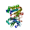
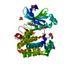
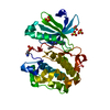
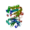
| Title | Human pepsin 3A in complex with a phosphonate inhibitor IVA-VAL-VAL-LEU(P)-(O)PHE-ALA-ALA-OME | ||||||
 Components Components | PEPSIN 3A | ||||||
 Keywords Keywords | HYDROLASE/HYDROLASE INHIBITOR / ASPARTIC PROTEINASE / PHOSPHONATE INHIBITOR / TRANSITION STATE ANALOGUE / HYDROLASE-HYDROLASE INHIBITOR COMPLEX | ||||||
| Function / homology |  Function and homology information Function and homology informationmultivesicular body lumen / pepsin A / Surfactant metabolism / digestion / aspartic-type endopeptidase activity / proteolysis / extracellular exosome Similarity search - Function | ||||||
| Biological species |  Homo sapiens (human) Homo sapiens (human) | ||||||
| Method |  X-RAY DIFFRACTION / Resolution: 1.96 Å X-RAY DIFFRACTION / Resolution: 1.96 Å | ||||||
 Authors Authors | Fujinaga, M. / Cherney, M.M. / Tarasova, N.I. / Bartlett, P.A. / Hanson, J.E. / James, M.N.G. | ||||||
 Citation Citation | Journal: Acta Crystallogr.,Sect.D / Year: 2000 Title: Structural study of the complex between human pepsin and a phosphorus-containing peptidic -transition-state analog. Authors: Fujinaga, M. / Cherney, M.M. / Tarasova, N.I. / Bartlett, P.A. / Hanson, J.E. / James, M.N. #1: Journal: Protein Sci. / Year: 1995Title: Crystal structure of human pepsin and its complex with pepstatin Authors: Fujinaga, M. / Chernaia, M.M. / Tarasova, N.I. / Mosimann, S.C. / James, M.N.G. | ||||||
| History |
|
- Structure visualization
| Structure viewer | Molecule: MolmilJmol/JSmol |
|---|
- Downloads & links
-Download
| PDBx/mmCIF format | 1qrp.cif.gz | 72.9 KB | Display | PDBx/mmCIF format |
|---|---|---|---|---|
| PDB format | pdb1qrp.ent.gz | 58 KB | Display | PDB format |
| PDBx/mmJSON format | 1qrp.json.gz | Tree view | PDBx/mmJSON format | |
| Others |  Other downloads Other downloads |
-Validation report
| Summary document | 1qrp_validation.pdf.gz | 711 KB | Display | wwPDB validaton report |
|---|---|---|---|---|
| Full document | 1qrp_full_validation.pdf.gz | 712.7 KB | Display | |
| Data in XML | 1qrp_validation.xml.gz | 15.2 KB | Display | |
| Data in CIF | 1qrp_validation.cif.gz | 21.6 KB | Display | |
| Arichive directory | https://data.pdbj.org/pub/pdb/validation_reports/qr/1qrpftp://data.pdbj.org/pub/pdb/validation_reports/qr/1qrp | HTTPS FTP |
-Related structure data
| Similar structure data |
|---|


-Links
 PDBj
PDBj
- Assembly
| Deposited unit | 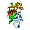
| ||||||||
|---|---|---|---|---|---|---|---|---|---|
| 1 |
| ||||||||
| Unit cell |
|
-Components
| #1: Protein | Mass: 34631.887 Da / Num. of mol.: 1 / Source method: isolated from a natural source / Source: (natural) Homo sapiens (human) / References: UniProt: P00790, UniProt: P0DJD7*PLUS |
|---|---|
| #2: Chemical | ChemComp-HH0 /   Type: peptide-like, Peptide-like / Class: Inhibitor / Mass: 753.863 Da / Num. of mol.: 1 / Source method: obtained synthetically / Formula: C36H60N5O10P / References: IVA VAL VAL P0L ALA MA Type: peptide-like, Peptide-like / Class: Inhibitor / Mass: 753.863 Da / Num. of mol.: 1 / Source method: obtained synthetically / Formula: C36H60N5O10P / References: IVA VAL VAL P0L ALA MA |
| #3: Water | ChemComp-HOH /  Mass: 18.015 Da / Num. of mol.: 135 / Source method: isolated from a natural source / Formula: H2O Mass: 18.015 Da / Num. of mol.: 135 / Source method: isolated from a natural source / Formula: H2O |
-Experimental details
-Experiment
| Experiment | Method: X-RAY DIFFRACTION / Number of used crystals: 1 |
|---|
- Sample preparation
| Crystal | Density Matthews: 3.14 Å3/Da / Density % sol: 60.77 % | ||||||||||||||||||||||||||||||||||||
|---|---|---|---|---|---|---|---|---|---|---|---|---|---|---|---|---|---|---|---|---|---|---|---|---|---|---|---|---|---|---|---|---|---|---|---|---|---|
| Crystal grow | Temperature: 293 K / Method: vapor diffusion, hanging drop / pH: 5 Details: 40% AMMONIUM SULFATE, 100MM SODIUM ACETATE, 5% MPD, pH 5.0, VAPOR DIFFUSION, HANGING DROP, temperature 293K | ||||||||||||||||||||||||||||||||||||
| Crystal grow | *PLUS Method: vapor diffusion | ||||||||||||||||||||||||||||||||||||
| Components of the solutions | *PLUS
|
-Data collection
| Diffraction | Mean temperature: 293 K |
|---|---|
| Diffraction source | Source: ROTATING ANODE / Type: RIGAKU RU200 / Wavelength: 1.5418 |
| Detector | Type: UCSD MARK III / Detector: AREA DETECTOR / Date: Aug 29, 1994 |
| Radiation | Protocol: SINGLE WAVELENGTH / Monochromatic (M) / Laue (L): M / Scattering type: x-ray |
| Radiation wavelength | Wavelength: 1.5418 Å / Relative weight: 1 |
| Reflection | Resolution: 1.96→65 Å / Num. all: 31990 / % possible obs: 89.3 % / Observed criterion σ(F): 3 / Redundancy: 5.9 % / Rmerge(I) obs: 0.108 |
| Reflection shell | Resolution: 1.96→2 Å / % possible all: 64.2 |
| Reflection | *PLUS Num. obs: 31990 / Num. measured all: 188324 |
| Reflection shell | *PLUS % possible obs: 64.2 % |
- Processing
| Software |
| ||||||||||||
|---|---|---|---|---|---|---|---|---|---|---|---|---|---|
| Refinement | Resolution: 1.96→30 Å / σ(F): 0 / σ(I): 0 / Stereochemistry target values: GROMOS87 FORCE FIELD
| ||||||||||||
| Refinement step | Cycle: LAST / Resolution: 1.96→30 Å
| ||||||||||||
| Refine LS restraints |
| ||||||||||||
| Software | *PLUS Name: GROMOS87 / Classification: refinement | ||||||||||||
| Refinement | *PLUS Lowest resolution: 30 Å / σ(F): 0 / Rfactor obs: 0.2 | ||||||||||||
| Solvent computation | *PLUS | ||||||||||||
| Displacement parameters | *PLUS |
