 Movie
Movie Controller


+ Open data

- Basic information
| Entry | Database: PDB / ID: 1qp1 | ||||||
|---|---|---|---|---|---|---|---|
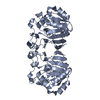
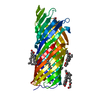
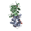
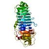
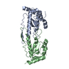
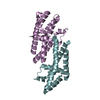
| Title | KAPPA VARIABLE LIGHT CHAIN | ||||||
 Components Components | BENCE-JONES KAPPA I ANTIBODY BRE (LIGHT CHAIN) | ||||||
 Keywords Keywords | IMMUNE SYSTEM / BETA SANDWICH / DOUBLE SPIRAL | ||||||
| Function / homology |  Function and homology information Function and homology informationCD22 mediated BCR regulation / Fc epsilon receptor (FCERI) signaling / Classical antibody-mediated complement activation / Initial triggering of complement / FCGR activation / Role of LAT2/NTAL/LAB on calcium mobilization / Role of phospholipids in phagocytosis / immunoglobulin complex / Scavenging of heme from plasma / antigen binding ...CD22 mediated BCR regulation / Fc epsilon receptor (FCERI) signaling / Classical antibody-mediated complement activation / Initial triggering of complement / FCGR activation / Role of LAT2/NTAL/LAB on calcium mobilization / Role of phospholipids in phagocytosis / immunoglobulin complex / Scavenging of heme from plasma / antigen binding / FCERI mediated Ca+2 mobilization / FCGR3A-mediated IL10 synthesis / Regulation of Complement cascade / Antigen activates B Cell Receptor (BCR) leading to generation of second messengers / Cell surface interactions at the vascular wall / FCGR3A-mediated phagocytosis / FCERI mediated MAPK activation / Regulation of actin dynamics for phagocytic cup formation / Immunoregulatory interactions between a Lymphoid and a non-Lymphoid cell / FCERI mediated NF-kB activation / blood microparticle / Potential therapeutics for SARS / adaptive immune response / immune response / extracellular region / identical protein binding / plasma membrane Similarity search - Function | ||||||
| Biological species |  Homo sapiens (human) Homo sapiens (human) | ||||||
| Method |  X-RAY DIFFRACTION / MOLECULAR REPLACEMENT / Resolution: 2.06 Å X-RAY DIFFRACTION / MOLECULAR REPLACEMENT / Resolution: 2.06 Å | ||||||
 Authors Authors | Steinrauf, L.K. | ||||||
 Citation Citation | Journal: J.Biochem.(Tokyo) / Year: 1999 Title: Molecular structure of the amyloid-forming protein kappa I Bre. Authors: Steinrauf, L.K. / Chiang, M.Y. / Shiuan, D. #1: Journal: Am.Cryst.Assoc.,Abstr.Papers (Annual Meeting) / Year: 1993Title: Structure of an Immunoglubulin Kappa Variable Light Chain Associated with Primary Amyloidosis Authors: Steinrauf, L.K. / Hamilton, J.A. / Clawson, D. / Liepnieks, J. / Murrell, J. / Benson, M.D. #2: Journal: Proc.Natl.Acad.Sci.USA / Year: 1995Title: Tertiary Structure of an Amyloid Immunoglobulin Light Chain Protein: A Proposed Model for Amyloid Fibril Formation Authors: Schormann, N. / Murrell, J.R. / Liepnieks, J.R. / Benson, M.D. | ||||||
| History |
|
- Structure visualization
| Structure viewer | Molecule: MolmilJmol/JSmol |
|---|
- Downloads & links
-Download
| PDBx/mmCIF format | 1qp1.cif.gz | 76.8 KB | Display | PDBx/mmCIF format |
|---|---|---|---|---|
| PDB format | pdb1qp1.ent.gz | 58.6 KB | Display | PDB format |
| PDBx/mmJSON format | 1qp1.json.gz | Tree view | PDBx/mmJSON format | |
| Others |  Other downloads Other downloads |
-Validation report
| Arichive directory | https://data.pdbj.org/pub/pdb/validation_reports/qp/1qp1ftp://data.pdbj.org/pub/pdb/validation_reports/qp/1qp1 | HTTPS FTP |
|---|
-Related structure data
| Related structure data | |
|---|---|
| Similar structure data |


-Links
 PDBj
PDBj


- Assembly
| Deposited unit | 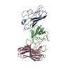
| ||||||||
|---|---|---|---|---|---|---|---|---|---|
| 1 |
| ||||||||
| Unit cell |
| ||||||||
| Components on special symmetry positions |
|
-Components
| #1: Antibody | Mass: 11787.930 Da / Num. of mol.: 3 / Fragment: IMMUNOGLOBULIN FRAGMENT, VARIABLE DOMAIN Source method: isolated from a genetically manipulated source Source: (gene. exp.) Homo sapiens (human) / Tissue: BONE MARROW / Plasmid: PCZ11 / Production host:  #2: Water | ChemComp-HOH / |  Mass: 18.015 Da / Num. of mol.: 191 / Source method: isolated from a natural source / Formula: H2O Mass: 18.015 Da / Num. of mol.: 191 / Source method: isolated from a natural source / Formula: H2OHas protein modification | Y | |
|---|
-Experimental details
-Experiment
| Experiment | Method: X-RAY DIFFRACTION / Number of used crystals: 1 |
|---|
- Sample preparation
| Crystal | Density Matthews: 3.26 Å3/Da / Density % sol: 62.24 % | ||||||||||||||||||||
|---|---|---|---|---|---|---|---|---|---|---|---|---|---|---|---|---|---|---|---|---|---|
| Crystal grow | Temperature: 300 K / Method: vapor diffusion, hanging drop / pH: 5.4 Details: AMMONIUM SULFATE, CITRATE BUFFER, pH 5.4, VAPOR DIFFUSION, HANGING DROP, temperature 300K | ||||||||||||||||||||
| Crystal grow | *PLUS Method: unknown | ||||||||||||||||||||
| Components of the solutions | *PLUS
|
-Data collection
| Diffraction | Mean temperature: 293 K |
|---|---|
| Diffraction source | Source: ROTATING ANODE / Type: RIGAKU RU300 / Wavelength: 1.5418 |
| Detector | Type: RIGAKU RAXIS II / Detector: IMAGE PLATE / Date: Aug 11, 1992 |
| Radiation | Monochromator: BENT CRYSTAL / Protocol: SINGLE WAVELENGTH / Monochromatic (M) / Laue (L): M / Scattering type: x-ray |
| Radiation wavelength | Wavelength: 1.5418 Å / Relative weight: 1 |
| Reflection | Resolution: 2.06→60 Å / Num. all: 89999 / Num. obs: 22485 / % possible obs: 68.9 % / Observed criterion σ(F): 1 / Observed criterion σ(I): 1 / Redundancy: 5 % / Biso Wilson estimate: 24 Å2 / Rmerge(I) obs: 0.12 / Net I/σ(I): 24 |
| Reflection shell | Resolution: 2.06→2.18 Å / Redundancy: 2 % / Rmerge(I) obs: 0.2 / Mean I/σ(I) obs: 1.92 / % possible all: 50.25 |
| Reflection shell | *PLUS % possible obs: 50.2 % |
- Processing
| Software |
| |||||||||||||||||||||||||||||||||
|---|---|---|---|---|---|---|---|---|---|---|---|---|---|---|---|---|---|---|---|---|---|---|---|---|---|---|---|---|---|---|---|---|---|---|
| Refinement | Method to determine structure: MOLECULAR REPLACEMENT Starting model: THE BENCE-JONES PROTEINS ROY: COLMAN, SCHRAMM, GURS J. MOL. BIOL. 116, 73, 1977 AND REI: EPP, COLMAN, FEHLHAMMER, BODE, SCHIFFER, HUBER, AND PALM EUR. J. BIOCHEM. 45, 513 1974 Resolution: 2.06→20 Å / Num. parameters: 1040 / Num. restraintsaints: 984 / Cross valid method: FREE R / σ(F): 4 / σ(I): 2 / Stereochemistry target values: ENGH AND HUBER
| |||||||||||||||||||||||||||||||||
| Refine analyze | Occupancy sum hydrogen: 0 / Occupancy sum non hydrogen: 2708 | |||||||||||||||||||||||||||||||||
| Refinement step | Cycle: LAST / Resolution: 2.06→20 Å
| |||||||||||||||||||||||||||||||||
| Refine LS restraints |
| |||||||||||||||||||||||||||||||||
| Software | *PLUS Name: SHELXL-97 / Classification: refinement | |||||||||||||||||||||||||||||||||
| Refine LS restraints | *PLUS
|
