- PDB-4wa0: The structure of a possible adhesin C-terminal domain from Caldic... -
+
Open data
ID or keywords:
Loading...
-
Basic information
Entry
Database: PDB / ID: 4wa0
Title
The structure of a possible adhesin C-terminal domain from Caldicellulosiruptor kronotskyensis
Components
possible adhesin
Keywords
CELL ADHESION / Beta-helix / Caldicellulosiruptor
Function / homology
Autotransporter, pectate lyase C-like domain superfamily / membrane / metal ion binding / Type 4 fimbrial biogenesis protein PilX N-terminal domain-containing protein
Mass: 18.015 Da / Num. of mol.: 549 / Source method: isolated from a natural source / Formula: H2O
Has protein modification
Y
Sequence details
THIS MOLECULE WAS PRODUCED WITH THERMOLYSIN CLEAVAGE FROM A LARGER PROTEIN. THE EXACT SEQUENCE OF ...THIS MOLECULE WAS PRODUCED WITH THERMOLYSIN CLEAVAGE FROM A LARGER PROTEIN. THE EXACT SEQUENCE OF THE CRYSTALLIZED ENTITY IS UNKNOWN.
-
Experimental details
-
Experiment
Experiment
Method: X-RAY DIFFRACTION
-
Sample preparation
Crystal
Density Matthews: 2.34 Å3/Da / Density % sol: 47.35 %
Crystal grow
Temperature: 294 K / Method: vapor diffusion, sitting drop / pH: 3.4 Details: PEG ion HT screen from Hampton Research (Aliso Viejo, CA) condition G12 containing 0.07 M Citric acid, 0.03 M BIS-TRIS propane, and 16% (w/v) Polyethylene glycol 3350
-
Data collection
Diffraction
Mean temperature: 100 K
Diffraction source
Source: ROTATING ANODE / Type: BRUKER AXS MICROSTAR / Wavelength: 1.5418 Å
Monochromator: HELIOS MIRRORS / Protocol: SINGLE WAVELENGTH / Monochromatic (M) / Laue (L): M / Scattering type: x-ray
Radiation wavelength
Wavelength: 1.5418 Å / Relative weight: 1
Reflection
Resolution: 1.7→55 Å / Num. obs: 40244 / % possible obs: 99.8 % / Redundancy: 8.24 % / Biso Wilson estimate: 14.323 Å2 / Net I/σ(I): 17.48
Reflection shell
Resolution: 1.7→1.8 Å / Redundancy: 5.69 % / Mean I/σ(I) obs: 2.36 / % possible all: 98.8
-
Processing
Software
Name
Version
Classification
PROTEUM PLUS
3013.8-1
datascaling
Coot
modelbuilding
REFMAC
5.8.0073
refinement
Refinement
Method to determine structure: SAD / Resolution: 1.7→53.98 Å / Cor.coef. Fo:Fc: 0.972 / Cor.coef. Fo:Fc free: 0.956 / SU B: 2.281 / SU ML: 0.074 / Cross valid method: THROUGHOUT / ESU R: 0.099 / ESU R Free: 0.102 / Stereochemistry target values: MAXIMUM LIKELIHOOD / Details: HYDROGENS HAVE BEEN ADDED IN THE RIDING POSITIONS
Rfactor
Num. reflection
% reflection
Selection details
Rfree
0.1941
1968
4.9 %
RANDOM
Rwork
0.15
-
-
-
obs
0.1522
38105
99.53 %
-
Solvent computation
Ion probe radii: 0.8 Å / Shrinkage radii: 0.8 Å / VDW probe radii: 1.2 Å / Solvent model: MASK
 Movie
Movie Controller

 Yorodumi
 Open data
 Basic information
 Components
 Keywords
 Function and homology information
 Caldicellulosiruptor kronotskyensis (bacteria)
 X-RAY DIFFRACTION /
  Authors
 Citation
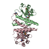
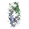
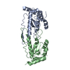
 Structure visualization
 Downloads & links
 Other downloads
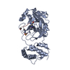


 PDBj
PDBj Assembly
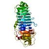

 Mass: 24.305 Da / Num. of mol.: 1 / Source method: obtained synthetically / Formula: Mg
 Mass: 18.015 Da / Num. of mol.: 549 / Source method: isolated from a natural source / Formula: H2O
 Sample preparation
 Processing