 Movie
Movie Controller

[English] 日本語
 Yorodumi
- PDB-6iru: Crystal structure of Peptidase E from Deinococcus radiodurans in ... -
+ Open data

- Basic information
| Entry | Database: PDB / ID: 6iru | ||||||
|---|---|---|---|---|---|---|---|
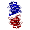
| Title | Crystal structure of Peptidase E from Deinococcus radiodurans in P6422 space group | ||||||
 Components Components | peptidase DR_1070 | ||||||
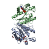
 Keywords Keywords | HYDROLASE / S51 peptidase / peptidase E / dimer / active site / esterase | ||||||
| Function / homology | Peptidase S51 / Peptidase family S51 / Class I glutamine amidotransferase-like / Hydrolases; Acting on peptide bonds (peptidases); Serine endopeptidases / serine-type peptidase activity / proteolysis / Uncharacterized peptidase DR_1070 Function and homology information Function and homology information | ||||||
| Biological species |  Deinococcus radiodurans (radioresistant) Deinococcus radiodurans (radioresistant) | ||||||
| Method |  X-RAY DIFFRACTION / SYNCHROTRON / MOLECULAR REPLACEMENT / Resolution: 2.7 Å X-RAY DIFFRACTION / SYNCHROTRON / MOLECULAR REPLACEMENT / Resolution: 2.7 Å | ||||||
 Authors Authors | Yadav, P. / Chandravanshi, K. / Kumar, A. / Makde, R.D. | ||||||
 Citation Citation | Journal: Proteins / Year: 2019 Title: Catalytic triad heterogeneity in S51 peptidase family: Structural basis for functional variability. Authors: Yadav, P. / Goyal, V.D. / Chandravanshi, K. / Kumar, A. / Gokhale, S.M. / Jamdar, S.N. / Makde, R.D. | ||||||
| History |
|
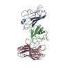
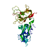
- Structure visualization
| Structure viewer | Molecule: MolmilJmol/JSmol |
|---|
- Downloads & links
-Download
| PDBx/mmCIF format | 6iru.cif.gz | 229.7 KB | Display | PDBx/mmCIF format |
|---|---|---|---|---|
| PDB format | pdb6iru.ent.gz | 186 KB | Display | PDB format |
| PDBx/mmJSON format | 6iru.json.gz | Tree view | PDBx/mmJSON format | |
| Others |  Other downloads Other downloads |
-Validation report
| Arichive directory | https://data.pdbj.org/pub/pdb/validation_reports/ir/6iruftp://data.pdbj.org/pub/pdb/validation_reports/ir/6iru | HTTPS FTP |
|---|
-Related structure data
| Related structure data | 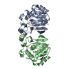 6a4tSC S: Starting model for refinement C: citing same article ( |
|---|---|
| Similar structure data |


-Links
 PDBj
PDBj

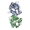
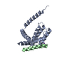
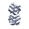
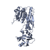
- Assembly
| Deposited unit | 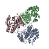
| ||||||||
|---|---|---|---|---|---|---|---|---|---|
| 1 | 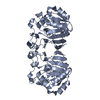
| ||||||||
| 2 | 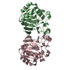
| ||||||||
| Unit cell |
|
-Components
| #1: Protein | Mass: 22729.799 Da / Num. of mol.: 3 Source method: isolated from a genetically manipulated source Source: (gene. exp.) Deinococcus radiodurans (radioresistant)Strain: ATCC 13939 / DSM 20539 / JCM 16871 / LMG 4051 / NBRC 15346 / NCIMB 9279 / R1 / VKM B-1422 Gene: DR_1070 / Plasmid: pet28a / Production host: References: UniProt: Q9RVF9, Hydrolases; Acting on peptide bonds (peptidases); Serine endopeptidases #2: Water | ChemComp-HOH / |  Mass: 18.015 Da / Num. of mol.: 8 / Source method: isolated from a natural source / Formula: H2O Mass: 18.015 Da / Num. of mol.: 8 / Source method: isolated from a natural source / Formula: H2O |
|---|
-Experimental details
-Experiment
| Experiment | Method: X-RAY DIFFRACTION / Number of used crystals: 1 |
|---|
- Sample preparation
| Crystal | Density Matthews: 2.69 Å3/Da / Density meas: 54.32 Mg/m3 / Density % sol: 58.57 % / Description: hexagonal shape (200-250 micron) |
|---|---|
| Crystal grow | Temperature: 294 K / Method: microbatch / pH: 6.5 / Details: 0.1 M Bis-tris pH 6.5, 25 % PEG3350 / PH range: 5.5-7.5 |
-Data collection
| Diffraction | Mean temperature: 100 K |
|---|---|
| Diffraction source | Source: SYNCHROTRON / Site: RRCAT INDUS-2  / Beamline: PX-BL21 / Wavelength: 0.9778 Å / Beamline: PX-BL21 / Wavelength: 0.9778 Å |
| Detector | Type: MAR scanner 345 mm plate / Detector: IMAGE PLATE / Date: Sep 15, 2018 / Details: mirrors |
| Radiation | Monochromator: Si111 / Protocol: SINGLE WAVELENGTH / Monochromatic (M) / Laue (L): M / Scattering type: x-ray |
| Radiation wavelength | Wavelength: 0.9778 Å / Relative weight: 1 |
| Reflection | Resolution: 2.7→48.14 Å / Num. obs: 23227 / % possible obs: 100 % / Redundancy: 14.5 % / Biso Wilson estimate: 58.8 Å2 / CC1/2: 0.999 / Rmerge(I) obs: 0.107 / Rpim(I) all: 0.028 / Rrim(I) all: 0.11 / Net I/σ(I): 23.5 |
| Reflection shell | Resolution: 2.7→2.83 Å / Redundancy: 14.9 % / Rmerge(I) obs: 1.147 / Num. unique obs: 3029 / CC1/2: 0.909 / Rpim(I) all: 0.297 / Rrim(I) all: 1.186 / % possible all: 100 |
- Processing
| Software |
| |||||||||||||||||||||||||||||||||||||||||||||||||||||||||||||||
|---|---|---|---|---|---|---|---|---|---|---|---|---|---|---|---|---|---|---|---|---|---|---|---|---|---|---|---|---|---|---|---|---|---|---|---|---|---|---|---|---|---|---|---|---|---|---|---|---|---|---|---|---|---|---|---|---|---|---|---|---|---|---|---|---|
| Refinement | Method to determine structure: MOLECULAR REPLACEMENT Starting model: 6A4T Resolution: 2.7→16.026 Å / SU ML: 0.33 / Cross valid method: FREE R-VALUE / σ(F): 1.34 / Phase error: 26.68
| |||||||||||||||||||||||||||||||||||||||||||||||||||||||||||||||
| Solvent computation | Shrinkage radii: 0.9 Å / VDW probe radii: 1.11 Å | |||||||||||||||||||||||||||||||||||||||||||||||||||||||||||||||
| Displacement parameters | Biso mean: 64 Å2 | |||||||||||||||||||||||||||||||||||||||||||||||||||||||||||||||
| Refinement step | Cycle: LAST / Resolution: 2.7→16.026 Å
| |||||||||||||||||||||||||||||||||||||||||||||||||||||||||||||||
| Refine LS restraints |
| |||||||||||||||||||||||||||||||||||||||||||||||||||||||||||||||
| LS refinement shell |
| |||||||||||||||||||||||||||||||||||||||||||||||||||||||||||||||
| Refinement TLS params. | Method: refined / Origin x: 17.4828 Å / Origin y: -67.2766 Å / Origin z: 11.1551 Å
| |||||||||||||||||||||||||||||||||||||||||||||||||||||||||||||||
| Refinement TLS group | Selection details: all |
