 Movie
Movie Controller

[English] 日本語
 Yorodumi
- PDB-1qh4: CRYSTAL STRUCTURE OF CHICKEN BRAIN-TYPE CREATINE KINASE AT 1.41 A... -
+ Open data

- Basic information
| Entry | Database: PDB / ID: 1qh4 | ||||||
|---|---|---|---|---|---|---|---|
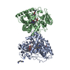
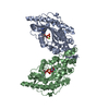
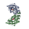
| Title | CRYSTAL STRUCTURE OF CHICKEN BRAIN-TYPE CREATINE KINASE AT 1.41 ANGSTROM RESOLUTION | ||||||
 Components Components | CREATINE KINASE | ||||||
 Keywords Keywords | TRANSFERASE / BRAIN-TYPE CREATINE KINASE / CANCER / CELLULAR ENERGY METABOLISM / GUANIDINO KINASE / NEURODEGENERATIVE DISORDERS | ||||||
| Function / homology |  Function and homology information Function and homology informationCreatine metabolism / RND3 GTPase cycle / extracellular membrane-bounded organelle / creatine kinase / phosphocreatine biosynthetic process / creatine kinase activity / ATP biosynthetic process / mitochondrion / : / ATP binding ...Creatine metabolism / RND3 GTPase cycle / extracellular membrane-bounded organelle / creatine kinase / phosphocreatine biosynthetic process / creatine kinase activity / ATP biosynthetic process / mitochondrion / : / ATP binding / plasma membrane / cytosol Similarity search - Function | ||||||
| Biological species |  | ||||||
| Method |  X-RAY DIFFRACTION / SYNCHROTRON / MOLECULAR REPLACEMENT / Resolution: 1.41 Å X-RAY DIFFRACTION / SYNCHROTRON / MOLECULAR REPLACEMENT / Resolution: 1.41 Å | ||||||
 Authors Authors | Eder, M. / Schlattner, U. / Becker, A. / Wallimann, T. / Kabsch, W. / Fritz-Wolf, K. | ||||||
 Citation Citation | Journal: Protein Sci. / Year: 1999 Title: Crystal structure of brain-type creatine kinase at 1.41 A resolution. Authors: Eder, M. / Schlattner, U. / Becker, A. / Wallimann, T. / Kabsch, W. / Fritz-Wolf, K. #1: Journal: Proc.Natl.Acad.Sci.USA / Year: 1998Title: Transition State Structure of Arginine Kinase: Implications for Catalysis of Bimolecular Reactions Authors: Zhou, G. / Somasundaram, T. / Blanc, E. / Parthasarathy, G. / Ellington, W.R. / Chapman, M.S. #2: Journal: Nature / Year: 1996Title: Structure of Mitochondrial Creatine Kinase. Authors: Fritz-Wolf, K. / Schnyder, T. / Wallimann, T. / Kabsch, W. | ||||||
| History |
|
- Structure visualization
| Structure viewer | Molecule: MolmilJmol/JSmol |
|---|
- Downloads & links
-Download
| PDBx/mmCIF format | 1qh4.cif.gz | 668.4 KB | Display | PDBx/mmCIF format |
|---|---|---|---|---|
| PDB format | pdb1qh4.ent.gz | 551.4 KB | Display | PDB format |
| PDBx/mmJSON format | 1qh4.json.gz | Tree view | PDBx/mmJSON format | |
| Others |  Other downloads Other downloads |
-Validation report
| Arichive directory | https://data.pdbj.org/pub/pdb/validation_reports/qh/1qh4ftp://data.pdbj.org/pub/pdb/validation_reports/qh/1qh4 | HTTPS FTP |
|---|
-Related structure data
| Related structure data | 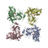 1crkS S: Starting model for refinement |
|---|---|
| Similar structure data |


-Links
 PDBj
PDBj

- Assembly
| Deposited unit | 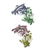
| ||||||||
|---|---|---|---|---|---|---|---|---|---|
| 1 | 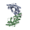
| ||||||||
| 2 | 
| ||||||||
| 3 | 
| ||||||||
| Unit cell |
|
-Components
| #1: Protein | Mass: 42799.523 Da / Num. of mol.: 4 / Fragment: B CHAIN Source method: isolated from a genetically manipulated source Source: (gene. exp.)  #2: Chemical | ChemComp-ACT /   Mass: 59.044 Da / Num. of mol.: 6 / Source method: obtained synthetically / Formula: C2H3O2 Mass: 59.044 Da / Num. of mol.: 6 / Source method: obtained synthetically / Formula: C2H3O2#3: Chemical | ChemComp-CA / |   Mass: 40.078 Da / Num. of mol.: 1 / Source method: obtained synthetically / Formula: Ca Mass: 40.078 Da / Num. of mol.: 1 / Source method: obtained synthetically / Formula: Ca#4: Water | ChemComp-HOH / |  Mass: 18.015 Da / Num. of mol.: 1467 / Source method: isolated from a natural source / Formula: H2O Mass: 18.015 Da / Num. of mol.: 1467 / Source method: isolated from a natural source / Formula: H2O |
|---|
-Experimental details
-Experiment
| Experiment | Method: X-RAY DIFFRACTION / Number of used crystals: 1 |
|---|
- Sample preparation
| Crystal | Density Matthews: 2.37 Å3/Da / Density % sol: 48 % | ||||||||||||||||||||||||||||||||||||||||||||||||
|---|---|---|---|---|---|---|---|---|---|---|---|---|---|---|---|---|---|---|---|---|---|---|---|---|---|---|---|---|---|---|---|---|---|---|---|---|---|---|---|---|---|---|---|---|---|---|---|---|---|
| Crystal grow | pH: 6.5 Details: 0.1M MES, 0.4M CA(OAC)2, 15% PE 2MM DTT, PH 6.5, 8 MG/ML PROTEIN | ||||||||||||||||||||||||||||||||||||||||||||||||
| Crystal | *PLUS Density % sol: 48 % | ||||||||||||||||||||||||||||||||||||||||||||||||
| Crystal grow | *PLUS Temperature: 20 ℃ / Method: vapor diffusion, sitting drop | ||||||||||||||||||||||||||||||||||||||||||||||||
| Components of the solutions | *PLUS
|
-Data collection
| Diffraction | Mean temperature: 100 K |
|---|---|
| Diffraction source | Source: SYNCHROTRON / Site: EMBL/DESY, HAMBURG  / Beamline: BW7B / Wavelength: 0.84 / Beamline: BW7B / Wavelength: 0.84 |
| Detector | Type: MAR scanner 345 mm plate / Detector: IMAGE PLATE / Date: Jul 22, 1998 / Details: BENT CRYSTAL |
| Radiation | Monochromator: GE SINGLE CRYSTAL / Protocol: SINGLE WAVELENGTH / Monochromatic (M) / Laue (L): M / Scattering type: x-ray |
| Radiation wavelength | Wavelength: 0.84 Å / Relative weight: 1 |
| Reflection | Resolution: 1.41→38 Å / Num. obs: 301191 / % possible obs: 99.1 % / Redundancy: 4 % / Rmerge(I) obs: 0.042 / Net I/σ(I): 17.2 |
| Reflection shell | Resolution: 1.41→1.5 Å / Redundancy: 3.5 % / Rmerge(I) obs: 0.215 / Mean I/σ(I) obs: 5 / % possible all: 97.1 |
| Reflection shell | *PLUS % possible obs: 97.1 % / Num. unique obs: 49959 |
- Processing
| Software |
| |||||||||||||||||||||||||||||||||
|---|---|---|---|---|---|---|---|---|---|---|---|---|---|---|---|---|---|---|---|---|---|---|---|---|---|---|---|---|---|---|---|---|---|---|
| Refinement | Method to determine structure: MOLECULAR REPLACEMENT Starting model: 1CRK, MITOCHONDRIAL CREATINE KINASE Resolution: 1.41→6 Å / Num. parameters: 12204 / Num. restraintsaints: 14868 / Cross valid method: FREE R / σ(F): 0 / Stereochemistry target values: ENGH AND HUBER Details: X-PLOR AND CNS WERE USED FOR INITIAL REFINEMENT. ANISOTROPIC REFINEMENT IN SHELX REDUCED FREE R (NO CUTOFF) BY 2.6% DISORDERED RESIDUES 321 - 330 (CHAIN A-D) WERE MODELED
| |||||||||||||||||||||||||||||||||
| Solvent computation | Solvent model: MOEWS & KRETSINGER, J.MOL.BIOL.91(1973)201-2 | |||||||||||||||||||||||||||||||||
| Refine analyze | Num. disordered residues: 15 / Occupancy sum hydrogen: 11644 / Occupancy sum non hydrogen: 13358 | |||||||||||||||||||||||||||||||||
| Refinement step | Cycle: LAST / Resolution: 1.41→6 Å
| |||||||||||||||||||||||||||||||||
| Refine LS restraints |
| |||||||||||||||||||||||||||||||||
| Software | *PLUS Name: SHELXL-97 / Classification: refinement | |||||||||||||||||||||||||||||||||
| Refinement | *PLUS Lowest resolution: 6 Å / σ(F): 0 / % reflection Rfree: 5.3 % / Rfactor obs: 0.134 / Rfactor Rwork: 0.1338 | |||||||||||||||||||||||||||||||||
| Solvent computation | *PLUS | |||||||||||||||||||||||||||||||||
| Displacement parameters | *PLUS | |||||||||||||||||||||||||||||||||
| Refine LS restraints | *PLUS
|
