 Movie
Movie Controller

[English] 日本語
 Yorodumi
- PDB-4v7n: Glycocyamine kinase, beta-beta homodimer from marine worm Namalyc... -
+ Open data

- Basic information
| Entry | Database: PDB / ID: 4v7n | |||||||||
|---|---|---|---|---|---|---|---|---|---|---|
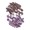
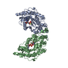
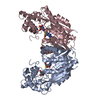
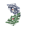
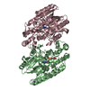
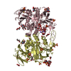
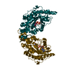
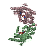
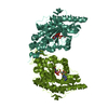
| Title | Glycocyamine kinase, beta-beta homodimer from marine worm Namalycastis sp., with transition state analog Mg(II)-ADP-NO3-glycocyamine. | |||||||||
 Components Components | Glycocyamine kinase beta chain | |||||||||
 Keywords Keywords | TRANSFERASE / phosphagen kinase / glycocyamine kinase / transition state analog / Kinase | |||||||||
| Function / homology |  Function and homology information Function and homology informationphosphocreatine biosynthetic process / creatine kinase activity / : / ATP binding Similarity search - Function | |||||||||
| Biological species |  Namalycastis sp. ST01 (invertebrata) Namalycastis sp. ST01 (invertebrata) | |||||||||
| Method |  X-RAY DIFFRACTION / SYNCHROTRON / MOLECULAR REPLACEMENT / Resolution: 2.3 Å X-RAY DIFFRACTION / SYNCHROTRON / MOLECULAR REPLACEMENT / Resolution: 2.3 Å | |||||||||
 Authors Authors | Lim, K. / Pullalarevu, S. / Herzberg, O. | |||||||||
 Citation Citation | Journal: Biochemistry / Year: 2010 Title: Structural basis for the mechanism and substrate specificity of glycocyamine kinase, a phosphagen kinase family member. Authors: Lim, K. / Pullalarevu, S. / Surabian, K.T. / Howard, A. / Suzuki, T. / Moult, J. / Herzberg, O. #1: Journal: COMP.BIOCHEM.PHYSIOL. B: BIOCHEM.MOL.BIOL. / Year: 2005Title: Isolation, characterization, and cDNA-derived amino acid sequence of glycocyamine kinase from the tropical marine worm Namalycastis sp. Authors: Mizuta, C. / Tanaka, K. / Suzuki, T. | |||||||||
| History |
|
- Structure visualization
| Structure viewer | Molecule: MolmilJmol/JSmol |
|---|
- Downloads & links
-Download
| PDBx/mmCIF format | 4v7n.cif.gz | 2.6 MB | Display | PDBx/mmCIF format |
|---|---|---|---|---|
| PDB format | pdb4v7n.ent.gz | Display | PDB format | |
| PDBx/mmJSON format | 4v7n.json.gz | Tree view | PDBx/mmJSON format | |
| Others |  Other downloads Other downloads |
-Validation report
| Arichive directory | https://data.pdbj.org/pub/pdb/validation_reports/v7/4v7nftp://data.pdbj.org/pub/pdb/validation_reports/v7/4v7n | HTTPS FTP |
|---|
-Related structure data
| Related structure data | 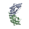 3l2dC 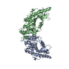 3l2eC  1u6rS  1vrpS C: citing same article ( S: Starting model for refinement |
|---|---|
| Similar structure data |


-Links
 PDBj
PDBj
- Assembly


-Components
-Protein , 1 types, 36 molecules AAABACADAEAFAGAHAIAJAKALAMANAOAPAQARBABBBCBDBEBFBGBHBIBJBKBL...
| #1: Protein | Mass: 44108.363 Da / Num. of mol.: 36 Source method: isolated from a genetically manipulated source Source: (gene. exp.) Namalycastis sp. ST01 (invertebrata) / Gene: GK-beta, GK_beta / Plasmid: pGK_beta221 / Production host:  |
|---|
-Non-polymers , 5 types, 4918 molecules 


| #2: Chemical | ChemComp-NMG /  Mass: 117.107 Da / Num. of mol.: 36 / Source method: obtained synthetically / Formula: C3H7N3O2 Mass: 117.107 Da / Num. of mol.: 36 / Source method: obtained synthetically / Formula: C3H7N3O2#3: Chemical | ChemComp-ADP /  Mass: 427.201 Da / Num. of mol.: 36 / Source method: obtained synthetically / Formula: C10H15N5O10P2 / Comment: ADP, energy-carrying molecule*YM Mass: 427.201 Da / Num. of mol.: 36 / Source method: obtained synthetically / Formula: C10H15N5O10P2 / Comment: ADP, energy-carrying molecule*YM#4: Chemical | ChemComp-MG /  Mass: 24.305 Da / Num. of mol.: 36 / Source method: obtained synthetically / Formula: Mg Mass: 24.305 Da / Num. of mol.: 36 / Source method: obtained synthetically / Formula: Mg#5: Chemical | ChemComp-NO3 /  Mass: 62.005 Da / Num. of mol.: 36 / Source method: obtained synthetically / Formula: NO3 Mass: 62.005 Da / Num. of mol.: 36 / Source method: obtained synthetically / Formula: NO3#6: Water | ChemComp-HOH / | Mass: 18.015 Da / Num. of mol.: 4774 / Source method: isolated from a natural source / Formula: H2O |
|---|
-Experimental details
-Experiment
| Experiment | Method: X-RAY DIFFRACTION / Number of used crystals: 1 |
|---|
- Sample preparation
| Crystal | Density Matthews: 2.27 Å3/Da / Density % sol: 45.9 % |
|---|---|
| Crystal grow | Temperature: 298 K / Method: vapor diffusion, hanging drop / pH: 8 Details: 22.5% polyethylene glycol monomethyl ether (PMME) 2000, 0.1M calcium acetate, 0.1M Tris HCl, pH 8, VAPOR DIFFUSION, HANGING DROP, temperature 298K |
-Data collection
| Diffraction | Mean temperature: 100 K |
|---|---|
| Diffraction source | Source: SYNCHROTRON / Site: APS  / Beamline: 22-ID / Wavelength: 1 Å / Beamline: 22-ID / Wavelength: 1 Å |
| Detector | Type: MARMOSAIC 300 mm CCD / Detector: CCD / Date: Nov 1, 2007 |
| Radiation | Protocol: SINGLE WAVELENGTH / Monochromatic (M) / Laue (L): M / Scattering type: x-ray |
| Radiation wavelength | Wavelength: 1 Å / Relative weight: 1 |
| Reflection | Resolution: 2.3→50 Å / Num. obs: 610182 / % possible obs: 96.9 % / Observed criterion σ(F): 0 / Observed criterion σ(I): 0 / Redundancy: 3.1 % / Rmerge(I) obs: 0.094 / Net I/σ(I): 13.8 |
| Reflection shell | Resolution: 2.3→2.38 Å / Redundancy: 2.3 % / Rmerge(I) obs: 0.403 / Mean I/σ(I) obs: 3.1 / % possible all: 81.2 |
- Processing
| Software |
| ||||||||||||||||
|---|---|---|---|---|---|---|---|---|---|---|---|---|---|---|---|---|---|
| Refinement | Method to determine structure: MOLECULAR REPLACEMENT Starting model: PDB entries 1u6r and 1vrp Resolution: 2.3→20 Å / Isotropic thermal model: isotropic / Cross valid method: THROUGHOUT / σ(F): 0 / σ(I): 0 / Stereochemistry target values: Engh & Huber Details: Used twinning refinement with twin operator (-h,-k,l) in Phenix program. Estimated twin fraction was 0.31.
| ||||||||||||||||
| Displacement parameters | Biso mean: 28 Å2 | ||||||||||||||||
| Refinement step | Cycle: LAST / Resolution: 2.3→20 Å
| ||||||||||||||||
| Refine LS restraints |
| ||||||||||||||||
| LS refinement shell | Resolution: 2.3→2.38 Å / Rfactor Rfree: 0.344 / Rfactor Rwork: 0.273 |
