 Movie
Movie Controller

[English] 日本語
 Yorodumi
- PDB-5foi: Crystal structure of mycinamicin VIII C21 methyl hydroxylase MycC... -
+ Open data

- Basic information
| Entry | Database: PDB / ID: 5foi | ||||||
|---|---|---|---|---|---|---|---|
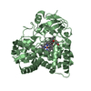
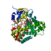
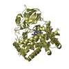
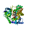
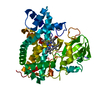
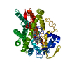
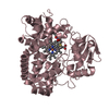
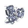
| Title | Crystal structure of mycinamicin VIII C21 methyl hydroxylase MycCI from Micromonospora griseorubida bound to mycinamicin VIII | ||||||
 Components Components | MYCINAMICIN VIII C21 METHYL HYDROXYLASE | ||||||
 Keywords Keywords | OXIDOREDUCTASE | ||||||
| Function / homology |  Function and homology information Function and homology informationOxidoreductases; Acting on paired donors, with incorporation or reduction of molecular oxygen / oxidoreductase activity, acting on paired donors, with incorporation or reduction of molecular oxygen / antibiotic biosynthetic process / monooxygenase activity / iron ion binding / heme binding Similarity search - Function | ||||||
| Biological species |  MICROMONOSPORA GRISEORUBIDA (bacteria) MICROMONOSPORA GRISEORUBIDA (bacteria) | ||||||
| Method |  X-RAY DIFFRACTION / SYNCHROTRON / MOLECULAR REPLACEMENT / Resolution: 2.21 Å X-RAY DIFFRACTION / SYNCHROTRON / MOLECULAR REPLACEMENT / Resolution: 2.21 Å | ||||||
 Authors Authors | Demars, M. / Sheng, F. / Podust, L.M. / Sherman, D.H. | ||||||
 Citation Citation | Journal: Acs Chem.Biol. / Year: 2016 Title: Biochemical and Structural Characterization of Mycci, a Versatile P450 Biocatalyst from the Mycinamicin Biosynthetic Pathway. Authors: Demars, M.D. / Sheng, F. / Park, S.R. / Lowell, A.N. / Podust, L.M. / Sherman, D.H. | ||||||
| History |
|
- Structure visualization
| Structure viewer | Molecule: MolmilJmol/JSmol |
|---|
- Downloads & links
-Download
| PDBx/mmCIF format | 5foi.cif.gz | 169.6 KB | Display | PDBx/mmCIF format |
|---|---|---|---|---|
| PDB format | pdb5foi.ent.gz | 132.7 KB | Display | PDB format |
| PDBx/mmJSON format | 5foi.json.gz | Tree view | PDBx/mmJSON format | |
| Others |  Other downloads Other downloads |
-Validation report
| Arichive directory | https://data.pdbj.org/pub/pdb/validation_reports/fo/5foiftp://data.pdbj.org/pub/pdb/validation_reports/fo/5foi | HTTPS FTP |
|---|
-Related structure data
| Related structure data |  3cv9S S: Starting model for refinement |
|---|---|
| Similar structure data |


-Links
 PDBj
PDBj

- Assembly
| Deposited unit | 
| ||||||||
|---|---|---|---|---|---|---|---|---|---|
| 1 | 
| ||||||||
| 2 | 
| ||||||||
| Unit cell |
|
-Components
-Protein , 1 types, 2 molecules AB
| #1: Protein | Mass: 45732.090 Da / Num. of mol.: 2 Source method: isolated from a genetically manipulated source Details: ENGINEERED SEQUENCE AT THE N-TERMINUS / Source: (gene. exp.) MICROMONOSPORA GRISEORUBIDA (bacteria) / Plasmid: PET28B / Production host: References: UniProt: Q83WF5, Oxidoreductases; Acting on paired donors, with incorporation or reduction of molecular oxygen |
|---|
-Non-polymers , 5 types, 156 molecules 


| #2: Chemical |  Mass: 616.487 Da / Num. of mol.: 2 / Source method: obtained synthetically / Formula: C34H32FeN4O4 Mass: 616.487 Da / Num. of mol.: 2 / Source method: obtained synthetically / Formula: C34H32FeN4O4#3: Chemical |  Mass: 507.702 Da / Num. of mol.: 2 / Source method: obtained synthetically / Formula: C29H49NO6 Mass: 507.702 Da / Num. of mol.: 2 / Source method: obtained synthetically / Formula: C29H49NO6#4: Chemical |  Mass: 92.094 Da / Num. of mol.: 2 / Source method: obtained synthetically / Formula: C3H8O3 Mass: 92.094 Da / Num. of mol.: 2 / Source method: obtained synthetically / Formula: C3H8O3#5: Chemical |  Mass: 145.246 Da / Num. of mol.: 3 / Source method: obtained synthetically / Formula: C7H19N3 Mass: 145.246 Da / Num. of mol.: 3 / Source method: obtained synthetically / Formula: C7H19N3#6: Water | ChemComp-HOH / | Mass: 18.015 Da / Num. of mol.: 147 / Source method: isolated from a natural source / Formula: H2O |
|---|
-Details
| Nonpolymer details | SPERMIDINE (SPD): PART OF CRYSTALLIZATION CONDITIONS MYCINAMICIN VIII (MY8): NATURAL SUBSTRATE OF ...SPERMIDINE |
|---|
-Experimental details
-Experiment
| Experiment | Method: X-RAY DIFFRACTION / Number of used crystals: 1 |
|---|
- Sample preparation
| Crystal | Density Matthews: 2.34 Å3/Da / Density % sol: 47.4 % / Description: NONE |
|---|---|
| Crystal grow | pH: 7 Details: 20% PEG 3350, 0.2 M CA ACETATE, 20 MM SPERMIDINE, 0.84 MM TCEP, pH 7 |
-Data collection
| Diffraction | Mean temperature: 110 K |
|---|---|
| Diffraction source | Source: SYNCHROTRON / Site: ALS  / Beamline: 8.3.1 / Wavelength: 1.11587 / Beamline: 8.3.1 / Wavelength: 1.11587 |
| Detector | Type: MARRESERCH / Detector: CCD / Date: Sep 3, 2015 / Details: MIRRORS |
| Radiation | Monochromator: SI(111) / Protocol: SINGLE WAVELENGTH / Monochromatic (M) / Laue (L): M / Scattering type: x-ray |
| Radiation wavelength | Wavelength: 1.11587 Å / Relative weight: 1 |
| Reflection | Resolution: 2.21→71 Å / Num. obs: 36326 / % possible obs: 91.1 % / Observed criterion σ(I): 0.5 / Redundancy: 2 % / Biso Wilson estimate: 31.6 Å2 / Rmerge(I) obs: 0.24 / Net I/σ(I): 3.8 |
| Reflection shell | Resolution: 2.21→2.33 Å / Redundancy: 1.9 % / Rmerge(I) obs: 1.47 / Mean I/σ(I) obs: 1.3 / % possible all: 71.2 |
- Processing
| Software |
| ||||||||||||||||||||||||||||||||||||||||||||||||||||||||||||||||||||||||||||||||||||||||||||||||||||||||||||||||||||||||||||||||||||||||||||||||||||||||||||||||||||||||||||||||||||||
|---|---|---|---|---|---|---|---|---|---|---|---|---|---|---|---|---|---|---|---|---|---|---|---|---|---|---|---|---|---|---|---|---|---|---|---|---|---|---|---|---|---|---|---|---|---|---|---|---|---|---|---|---|---|---|---|---|---|---|---|---|---|---|---|---|---|---|---|---|---|---|---|---|---|---|---|---|---|---|---|---|---|---|---|---|---|---|---|---|---|---|---|---|---|---|---|---|---|---|---|---|---|---|---|---|---|---|---|---|---|---|---|---|---|---|---|---|---|---|---|---|---|---|---|---|---|---|---|---|---|---|---|---|---|---|---|---|---|---|---|---|---|---|---|---|---|---|---|---|---|---|---|---|---|---|---|---|---|---|---|---|---|---|---|---|---|---|---|---|---|---|---|---|---|---|---|---|---|---|---|---|---|---|---|
| Refinement | Method to determine structure: MOLECULAR REPLACEMENT Starting model: PDB ENTRY 3CV9 Resolution: 2.21→71 Å / Cor.coef. Fo:Fc: 0.948 / Cor.coef. Fo:Fc free: 0.898 / SU B: 7.238 / SU ML: 0.178 / Cross valid method: THROUGHOUT / ESU R: 0.358 / ESU R Free: 0.251 / Stereochemistry target values: MAXIMUM LIKELIHOOD / Details: HYDROGENS HAVE BEEN ADDED IN THE RIDING POSITIONS
| ||||||||||||||||||||||||||||||||||||||||||||||||||||||||||||||||||||||||||||||||||||||||||||||||||||||||||||||||||||||||||||||||||||||||||||||||||||||||||||||||||||||||||||||||||||||
| Solvent computation | Ion probe radii: 0.8 Å / Shrinkage radii: 0.8 Å / VDW probe radii: 1.2 Å / Solvent model: MASK | ||||||||||||||||||||||||||||||||||||||||||||||||||||||||||||||||||||||||||||||||||||||||||||||||||||||||||||||||||||||||||||||||||||||||||||||||||||||||||||||||||||||||||||||||||||||
| Displacement parameters | Biso mean: 27.168 Å2
| ||||||||||||||||||||||||||||||||||||||||||||||||||||||||||||||||||||||||||||||||||||||||||||||||||||||||||||||||||||||||||||||||||||||||||||||||||||||||||||||||||||||||||||||||||||||
| Refinement step | Cycle: LAST / Resolution: 2.21→71 Å
| ||||||||||||||||||||||||||||||||||||||||||||||||||||||||||||||||||||||||||||||||||||||||||||||||||||||||||||||||||||||||||||||||||||||||||||||||||||||||||||||||||||||||||||||||||||||
| Refine LS restraints |
|