 Movie
Movie Controller

[English] 日本語
 Yorodumi
- PDB-3cv9: Crystal structure of vitamin D hydroxylase cytochrome P450 105A1 ... -
+ Open data

- Basic information
| Entry | Database: PDB / ID: 3cv9 | ||||||
|---|---|---|---|---|---|---|---|
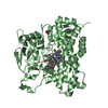
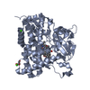
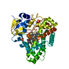
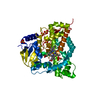
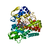
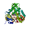
| Title | Crystal structure of vitamin D hydroxylase cytochrome P450 105A1 (R73A/R84A mutant) in complex with 1alpha,25-dihydroxyvitamin D3 | ||||||
 Components Components | Cytochrome P450-SU1 | ||||||
 Keywords Keywords | OXIDOREDUCTASE / P450 / BETA PRISM / HEME / IRON / METAL-BINDING / MONOOXYGENASE | ||||||
| Function / homology |  Function and homology information Function and homology informationvitamin D 1,25-hydroxylase / vitamin D3 metabolic process / oxidoreductase activity, acting on paired donors, with incorporation or reduction of molecular oxygen, NAD(P)H as one donor, and incorporation of one atom of oxygen / iron ion binding / heme binding / cytoplasm Similarity search - Function | ||||||
| Biological species |  Streptomyces griseolus (bacteria) Streptomyces griseolus (bacteria) | ||||||
| Method |  X-RAY DIFFRACTION / SYNCHROTRON / MOLECULAR REPLACEMENT / Resolution: 1.7 Å X-RAY DIFFRACTION / SYNCHROTRON / MOLECULAR REPLACEMENT / Resolution: 1.7 Å | ||||||
 Authors Authors | Hayashi, K. / Sugimoto, H. / Shinkyo, R. / Yamada, M. / Ikeda, S. / Ikushiro, S. / Kamakura, M. / Shiro, Y. / Sakaki, T. | ||||||
 Citation Citation | Journal: Biochemistry / Year: 2008 Title: Structure-based design of a highly active vitamin D hydroxylase from Streptomyces griseolus CYP105A1 Authors: Hayashi, K. / Sugimoto, H. / Shinkyo, R. / Yamada, M. / Ikeda, S. / Ikushiro, S. / Kamakura, M. / Shiro, Y. / Sakaki, T. #1: Journal: Biochemistry / Year: 2008Title: Crystal Structure of CYP105A1 (P450SU-1) in Complex with 1alpha,25-Dihydroxyvitamin D3 Authors: Sugimoto, H. / Shinkyo, R. / Hayashi, K. / Yoneda, S. / Yamada, M. / Kamakura, M. / Ikushiro, S. / Shiro, Y. / Sakaki, T. | ||||||
| History |
|
- Structure visualization
| Structure viewer | Molecule: MolmilJmol/JSmol |
|---|
- Downloads & links
-Download
| PDBx/mmCIF format | 3cv9.cif.gz | 105.5 KB | Display | PDBx/mmCIF format |
|---|---|---|---|---|
| PDB format | pdb3cv9.ent.gz | 78.9 KB | Display | PDB format |
| PDBx/mmJSON format | 3cv9.json.gz | Tree view | PDBx/mmJSON format | |
| Others |  Other downloads Other downloads |
-Validation report
| Arichive directory | https://data.pdbj.org/pub/pdb/validation_reports/cv/3cv9ftp://data.pdbj.org/pub/pdb/validation_reports/cv/3cv9 | HTTPS FTP |
|---|
-Related structure data
| Related structure data | 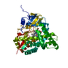 3cv8C  2zbzS S: Starting model for refinement C: citing same article ( |
|---|---|
| Similar structure data |


-Links
 PDBj
PDBj

- Assembly
| Deposited unit | 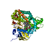
| ||||||||
|---|---|---|---|---|---|---|---|---|---|
| 1 |
| ||||||||
| Unit cell |
|
-Components
| #1: Protein | Mass: 44905.629 Da / Num. of mol.: 1 / Mutation: R73A, R84A Source method: isolated from a genetically manipulated source Source: (gene. exp.) Streptomyces griseolus (bacteria) / Strain: ATCC 11796 / Gene: CYP105A1 / Plasmid: PKK223-3 / Production host: |
|---|---|
| #2: Chemical | ChemComp-HEM /   Mass: 616.487 Da / Num. of mol.: 1 / Source method: obtained synthetically / Formula: C34H32FeN4O4 Mass: 616.487 Da / Num. of mol.: 1 / Source method: obtained synthetically / Formula: C34H32FeN4O4 |
| #3: Chemical | ChemComp-VDX /   Mass: 416.636 Da / Num. of mol.: 1 / Source method: obtained synthetically / Formula: C27H44O3 Mass: 416.636 Da / Num. of mol.: 1 / Source method: obtained synthetically / Formula: C27H44O3 |
| #4: Water | ChemComp-HOH /  Mass: 18.015 Da / Num. of mol.: 463 / Source method: isolated from a natural source / Formula: H2O Mass: 18.015 Da / Num. of mol.: 463 / Source method: isolated from a natural source / Formula: H2O |
| Sequence details | THERE IS A CONFLICT BETWEEN THE SEQUENCES GIVEN IN THE DEPOSITION AND THE DATABASE. ACCORDING TO ...THERE IS A CONFLICT BETWEEN THE SEQUENCES GIVEN IN THE DEPOSITION |
-Experimental details
-Experiment
| Experiment | Method: X-RAY DIFFRACTION / Number of used crystals: 1 |
|---|
- Sample preparation
| Crystal | Density Matthews: 2.24 Å3/Da / Density % sol: 45.07 % |
|---|---|
| Crystal grow | Temperature: 283 K / Method: vapor diffusion, sitting drop / pH: 6.2 Details: 26% PEGMME 2000, 0.1M Bis-tris, 0.2M sodium chlorife, pH 6.2, VAPOR DIFFUSION, SITTING DROP, temperature 283K |
-Data collection
| Diffraction | Mean temperature: 90 K |
|---|---|
| Diffraction source | Source: SYNCHROTRON / Site: SPring-8  / Beamline: BL44B2 / Wavelength: 1 Å / Beamline: BL44B2 / Wavelength: 1 Å |
| Detector | Type: ADSC QUANTUM 210 / Detector: CCD / Date: Jun 28, 2007 / Details: mirrors |
| Radiation | Monochromator: Si (1 1 1) Double crystal monochromator / Protocol: SINGLE WAVELENGTH / Monochromatic (M) / Laue (L): M / Scattering type: x-ray |
| Radiation wavelength | Wavelength: 1 Å / Relative weight: 1 |
| Reflection | Resolution: 1.7→20 Å / Num. all: 45139 / Num. obs: 45139 / % possible obs: 98.5 % / Observed criterion σ(I): -3 / Redundancy: 7.1 % / Biso Wilson estimate: 21.6 Å2 / Rsym value: 0.04 / Net I/σ(I): 28.8 |
| Reflection shell | Resolution: 1.7→1.76 Å / Redundancy: 5.3 % / Mean I/σ(I) obs: 5.1 / Num. unique all: 3913 / Rsym value: 0.337 / % possible all: 87.1 |
- Processing
| Software |
| ||||||||||||||||||||||||||||||||||||||||||||||||||||||||||||||||||||||||||||||||||||||||||
|---|---|---|---|---|---|---|---|---|---|---|---|---|---|---|---|---|---|---|---|---|---|---|---|---|---|---|---|---|---|---|---|---|---|---|---|---|---|---|---|---|---|---|---|---|---|---|---|---|---|---|---|---|---|---|---|---|---|---|---|---|---|---|---|---|---|---|---|---|---|---|---|---|---|---|---|---|---|---|---|---|---|---|---|---|---|---|---|---|---|---|---|
| Refinement | Method to determine structure: MOLECULAR REPLACEMENT Starting model: PDB ENTRY 2ZBZ Resolution: 1.7→20 Å / Cor.coef. Fo:Fc: 0.95 / Cor.coef. Fo:Fc free: 0.921 / SU B: 2.693 / SU ML: 0.091 / Cross valid method: THROUGHOUT / σ(F): 0 / ESU R: 0.126 / ESU R Free: 0.13 / Stereochemistry target values: MAXIMUM LIKELIHOOD
| ||||||||||||||||||||||||||||||||||||||||||||||||||||||||||||||||||||||||||||||||||||||||||
| Solvent computation | Ion probe radii: 0.8 Å / Shrinkage radii: 0.8 Å / VDW probe radii: 1.4 Å / Solvent model: MASK | ||||||||||||||||||||||||||||||||||||||||||||||||||||||||||||||||||||||||||||||||||||||||||
| Displacement parameters | Biso mean: 23.075 Å2
| ||||||||||||||||||||||||||||||||||||||||||||||||||||||||||||||||||||||||||||||||||||||||||
| Refinement step | Cycle: LAST / Resolution: 1.7→20 Å
| ||||||||||||||||||||||||||||||||||||||||||||||||||||||||||||||||||||||||||||||||||||||||||
| Refine LS restraints |
| ||||||||||||||||||||||||||||||||||||||||||||||||||||||||||||||||||||||||||||||||||||||||||
| LS refinement shell | Resolution: 1.7→1.744 Å / Total num. of bins used: 20
|
