 Movie
Movie Controller

[English] 日本語
 Yorodumi
- PDB-4wq0: Crystal structure of cytochrome P450 CYP107W1 from Streptomyces a... -
+ Open data

- Basic information
| Entry | Database: PDB / ID: 4wq0 | ||||||
|---|---|---|---|---|---|---|---|
| Title | Crystal structure of cytochrome P450 CYP107W1 from Streptomyces avermitilis in complex with Oligomycin A | ||||||
 Components Components | Cytochrome P450 | ||||||
 Keywords Keywords | OXIDOREDUCTASE/ANTIBIOTIC / P450 / CYP / CYP107W1 / Streptomyces avermitilis / oligomycin / OXIDOREDUCTASE-ANTIBIOTIC complex | ||||||
| Function / homology |  Function and homology information Function and homology informationoxidoreductase activity, acting on paired donors, with incorporation or reduction of molecular oxygen / monooxygenase activity / iron ion binding / heme binding Similarity search - Function | ||||||
| Biological species |  Streptomyces avermitilis (bacteria) Streptomyces avermitilis (bacteria) | ||||||
| Method |  X-RAY DIFFRACTION / SYNCHROTRON / SAD / Resolution: 2.7 Å X-RAY DIFFRACTION / SYNCHROTRON / SAD / Resolution: 2.7 Å | ||||||
| Model details | Biosynthesis of Oligomycin A | ||||||
 Authors Authors | Kang, L.W. / Kim, D.H. / Pham, T.V. / Han, S.H. | ||||||
 Citation Citation | Journal: To Be Published Title: Crystal structure of cytochrome P450 CYP107W1 from Streptomyces avermitilis in complex with Oligomycin A Authors: Kang, L.W. / Kim, D.H. / Pham, T.V. / Han, S.H. / Kim, J.H. / Lim, Y.R. / Park, H.G. / Cha, G.S. / Yun, C.H. / Chun, Y.J. | ||||||
| History |
|
- Structure visualization
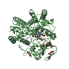
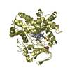
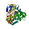
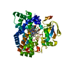
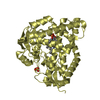
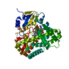
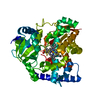
| Structure viewer | Molecule: MolmilJmol/JSmol |
|---|
- Downloads & links
-Download
| PDBx/mmCIF format | 4wq0.cif.gz | 95 KB | Display | PDBx/mmCIF format |
|---|---|---|---|---|
| PDB format | pdb4wq0.ent.gz | 70.5 KB | Display | PDB format |
| PDBx/mmJSON format | 4wq0.json.gz | Tree view | PDBx/mmJSON format | |
| Others |  Other downloads Other downloads |
-Validation report
| Arichive directory | https://data.pdbj.org/pub/pdb/validation_reports/wq/4wq0ftp://data.pdbj.org/pub/pdb/validation_reports/wq/4wq0 | HTTPS FTP |
|---|
-Related structure data
| Related structure data | |
|---|---|
| Similar structure data |


-Links
 PDBj
PDBj
- Assembly
| Deposited unit | 
| ||||||||
|---|---|---|---|---|---|---|---|---|---|
| 1 |
| ||||||||
| Unit cell |
| ||||||||
| Components on special symmetry positions |
| ||||||||
| Details | biological unit is the same as asym. |
-Components
| #1: Protein | Mass: 43314.551 Da / Num. of mol.: 1 / Fragment: UNP residues 8-404 Source method: isolated from a genetically manipulated source Source: (gene. exp.) Streptomyces avermitilis (bacteria) / Gene: olmB / Plasmid: pET 28a(+) / Production host: |
|---|---|
| #2: Chemical | ChemComp-HEM /   Mass: 616.487 Da / Num. of mol.: 1 / Source method: obtained synthetically / Formula: C34H32FeN4O4 Mass: 616.487 Da / Num. of mol.: 1 / Source method: obtained synthetically / Formula: C34H32FeN4O4 |
| #3: Chemical | ChemComp-EFO / 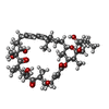  Mass: 791.062 Da / Num. of mol.: 1 / Source method: obtained synthetically / Formula: C45H74O11 Mass: 791.062 Da / Num. of mol.: 1 / Source method: obtained synthetically / Formula: C45H74O11 |
| #4: Water | ChemComp-HOH /  Mass: 18.015 Da / Num. of mol.: 15 / Source method: isolated from a natural source / Formula: H2O Mass: 18.015 Da / Num. of mol.: 15 / Source method: isolated from a natural source / Formula: H2O |
-Experimental details
-Experiment
| Experiment | Method: X-RAY DIFFRACTION / Number of used crystals: 1 |
|---|
- Sample preparation
| Crystal | Density Matthews: 3.61 Å3/Da / Density % sol: 65.93 % |
|---|---|
| Crystal grow | Temperature: 287 K / Method: vapor diffusion, hanging drop / pH: 6.5 / Details: Isopropanol, Ammonium citrate, citric acid |
-Data collection
| Diffraction | Mean temperature: 100 K |
|---|---|
| Diffraction source | Source: SYNCHROTRON / Site: PAL/PLS  / Beamline: 7A (6B, 6C1) / Wavelength: 0.979 Å / Beamline: 7A (6B, 6C1) / Wavelength: 0.979 Å |
| Detector | Type: ADSC QUANTUM 270 / Detector: CCD / Date: Dec 20, 2013 / Details: mirror |
| Radiation | Monochromator: DCM Si (111) Crystal / Protocol: SINGLE WAVELENGTH / Monochromatic (M) / Laue (L): M / Scattering type: x-ray |
| Radiation wavelength | Wavelength: 0.979 Å / Relative weight: 1 |
| Reflection | Resolution: 2.6→50 Å / Num. obs: 19627 / % possible obs: 99.6 % / Redundancy: 8.8 % / Net I/σ(I): 26.2 |
- Processing
| Software |
| |||||||||||||||||||||||||||||||||||||||||||||||||||||||||||||||||||||||||||
|---|---|---|---|---|---|---|---|---|---|---|---|---|---|---|---|---|---|---|---|---|---|---|---|---|---|---|---|---|---|---|---|---|---|---|---|---|---|---|---|---|---|---|---|---|---|---|---|---|---|---|---|---|---|---|---|---|---|---|---|---|---|---|---|---|---|---|---|---|---|---|---|---|---|---|---|---|
| Refinement | Method to determine structure: SAD / Resolution: 2.7→40.42 Å / Cor.coef. Fo:Fc: 0.947 / Cor.coef. Fo:Fc free: 0.907 / SU B: 19.374 / SU ML: 0.352 / Cross valid method: THROUGHOUT / σ(F): 0 / ESU R: 0.563 / ESU R Free: 0.355 / Stereochemistry target values: MAXIMUM LIKELIHOOD Details: HYDROGENS HAVE BEEN ADDED IN THE RIDING POSITIONS U VALUES : REFINED INDIVIDUALLY
| |||||||||||||||||||||||||||||||||||||||||||||||||||||||||||||||||||||||||||
| Solvent computation | Ion probe radii: 0.8 Å / Shrinkage radii: 0.8 Å / VDW probe radii: 1.2 Å / Solvent model: MASK | |||||||||||||||||||||||||||||||||||||||||||||||||||||||||||||||||||||||||||
| Displacement parameters | Biso max: 161.89 Å2 / Biso mean: 79.432 Å2 / Biso min: 47.14 Å2
| |||||||||||||||||||||||||||||||||||||||||||||||||||||||||||||||||||||||||||
| Refinement step | Cycle: final / Resolution: 2.7→40.42 Å
| |||||||||||||||||||||||||||||||||||||||||||||||||||||||||||||||||||||||||||
| Refine LS restraints |
| |||||||||||||||||||||||||||||||||||||||||||||||||||||||||||||||||||||||||||
| LS refinement shell | Resolution: 2.703→2.773 Å / Total num. of bins used: 20
|
