 Movie
Movie Controller

[English] 日本語
 Yorodumi
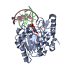
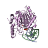
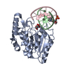
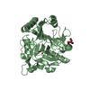
- PDB-1q3f: Uracil DNA glycosylase bound to a cationic 1-aza-2'-deoxyribose-c... -
+ Open data

- Basic information
| Entry | Database: PDB / ID: 1q3f | ||||||
|---|---|---|---|---|---|---|---|
| Title | Uracil DNA glycosylase bound to a cationic 1-aza-2'-deoxyribose-containing DNA | ||||||
 Components Components |
| ||||||
 Keywords Keywords | HYDROLASE/DNA / UDG / DNA repair / HYDROLASE-DNA COMPLEX | ||||||
| Function / homology |  Function and homology information Function and homology informationbase-excision repair, AP site formation via deaminated base removal / uracil-DNA glycosylase / depyrimidination / Displacement of DNA glycosylase by APEX1 / single strand break repair / uracil DNA N-glycosylase activity / isotype switching / ribosomal small subunit binding / somatic hypermutation of immunoglobulin genes / Recognition and association of DNA glycosylase with site containing an affected pyrimidine ...base-excision repair, AP site formation via deaminated base removal / uracil-DNA glycosylase / depyrimidination / Displacement of DNA glycosylase by APEX1 / single strand break repair / uracil DNA N-glycosylase activity / isotype switching / ribosomal small subunit binding / somatic hypermutation of immunoglobulin genes / Recognition and association of DNA glycosylase with site containing an affected pyrimidine / Cleavage of the damaged pyrimidine / Chromatin modifications during the maternal to zygotic transition (MZT) / base-excision repair / damaged DNA binding / negative regulation of apoptotic process / mitochondrion / nucleoplasm / nucleus Similarity search - Function | ||||||
| Biological species |  Homo sapiens (human) Homo sapiens (human) | ||||||
| Method |  X-RAY DIFFRACTION / MOLECULAR REPLACEMENT / Resolution: 1.9 Å X-RAY DIFFRACTION / MOLECULAR REPLACEMENT / Resolution: 1.9 Å | ||||||
 Authors Authors | Bianchet, M.A. / Seiple, L.A. / Jiang, Y.L. / Ichikawa, Y. / Amzel, L.M. / Stivers, J.T. | ||||||
 Citation Citation | Journal: Biochemistry / Year: 2003 Title: Electrostatic guidance of glycosyl cation migration along the reaction coordinate of uracil DNA glycosylase. Authors: Bianchet, M.A. / Seiple, L.A. / Jiang, Y.L. / Ichikawa, Y. / Amzel, L.M. / Stivers, J.T. | ||||||
| History |
| ||||||
| Remark 600 | HETEROGEN The NRI and URA is a transition state analog. This transition state analog has an uracil ...HETEROGEN The NRI and URA is a transition state analog. This transition state analog has an uracil and a modified deoxyribose that are not covalently connected to each other. |
- Structure visualization
| Structure viewer | Molecule: MolmilJmol/JSmol |
|---|
- Downloads & links
-Download
| PDBx/mmCIF format | 1q3f.cif.gz | 78.7 KB | Display | PDBx/mmCIF format |
|---|---|---|---|---|
| PDB format | pdb1q3f.ent.gz | 54.1 KB | Display | PDB format |
| PDBx/mmJSON format | 1q3f.json.gz | Tree view | PDBx/mmJSON format | |
| Others |  Other downloads Other downloads |
-Validation report
| Arichive directory | https://data.pdbj.org/pub/pdb/validation_reports/q3/1q3fftp://data.pdbj.org/pub/pdb/validation_reports/q3/1q3f | HTTPS FTP |
|---|
-Related structure data
| Related structure data |  1akxS S: Starting model for refinement |
|---|---|
| Similar structure data |


-Links
 PDBj
PDBj
- Assembly
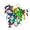
| Deposited unit | 
| ||||||||
|---|---|---|---|---|---|---|---|---|---|
| 1 |
| ||||||||
| Unit cell |
|
-Components
-DNA chain , 2 types, 2 molecules BC
| #1: DNA chain | Mass: 2586.712 Da / Num. of mol.: 1 / Source method: obtained synthetically / Details: 1-aza-2'-deoxyribose-containing DNA |
|---|---|
| #2: DNA chain | Mass: 3070.071 Da / Num. of mol.: 1 / Source method: obtained synthetically |
-Protein , 1 types, 1 molecules A
| #3: Protein | Mass: 25544.137 Da / Num. of mol.: 1 Source method: isolated from a genetically manipulated source Source: (gene. exp.) Homo sapiens (human) / Gene: UNG OR DGU OR UNG15 / Production host:  References: UniProt: P13051, Hydrolases; Glycosylases; Hydrolysing N-glycosyl compounds |
|---|
-Non-polymers , 3 types, 277 molecules 


| #4: Chemical | ChemComp-URA /  Mass: 112.087 Da / Num. of mol.: 1 / Source method: obtained synthetically / Formula: C4H4N2O2 Mass: 112.087 Da / Num. of mol.: 1 / Source method: obtained synthetically / Formula: C4H4N2O2 |
|---|---|
| #5: Chemical | ChemComp-PO4 /  Mass: 94.971 Da / Num. of mol.: 1 / Source method: obtained synthetically / Formula: PO4 Mass: 94.971 Da / Num. of mol.: 1 / Source method: obtained synthetically / Formula: PO4 |
| #6: Water | ChemComp-HOH / Mass: 18.015 Da / Num. of mol.: 275 / Source method: isolated from a natural source / Formula: H2O |
-Experimental details
-Experiment
| Experiment | Method: X-RAY DIFFRACTION / Number of used crystals: 1 |
|---|
- Sample preparation
| Crystal | Density Matthews: 2.54 Å3/Da / Density % sol: 51.5 % | ||||||||||||||||||||||||||||||||||||||||||||||||||||||
|---|---|---|---|---|---|---|---|---|---|---|---|---|---|---|---|---|---|---|---|---|---|---|---|---|---|---|---|---|---|---|---|---|---|---|---|---|---|---|---|---|---|---|---|---|---|---|---|---|---|---|---|---|---|---|---|
| Crystal grow | Temperature: 298 K / Method: vapor diffusion, hanging drop / pH: 6.5 Details: 18% PEG 4000, 10% dioxane, 100mM Hepes , pH 6.5, VAPOR DIFFUSION, HANGING DROP, temperature 298K | ||||||||||||||||||||||||||||||||||||||||||||||||||||||
| Components of the solutions |
| ||||||||||||||||||||||||||||||||||||||||||||||||||||||
| Crystal grow | *PLUS Method: vapor diffusion, hanging drop | ||||||||||||||||||||||||||||||||||||||||||||||||||||||
| Components of the solutions | *PLUS
|
-Data collection
| Diffraction | Mean temperature: 298 K |
|---|---|
| Diffraction source | Source: ROTATING ANODE / Type: RIGAKU RU300 / Wavelength: 1.5418 Å |
| Detector | Type: RIGAKU RAXIS IV / Detector: IMAGE PLATE / Date: Jan 22, 2003 / Details: mirrors |
| Radiation | Monochromator: mirrors / Protocol: SINGLE WAVELENGTH / Monochromatic (M) / Laue (L): M / Scattering type: x-ray |
| Radiation wavelength | Wavelength: 1.5418 Å / Relative weight: 1 |
| Reflection | Resolution: 1.9→36.51 Å / Num. obs: 24682 / % possible obs: 94.9 % / Observed criterion σ(F): 0 / Observed criterion σ(I): 0 / Redundancy: 4.7 % / Rsym value: 0.06 / Net I/σ(I): 16.2 |
| Reflection shell | Resolution: 1.9→2 Å / Redundancy: 2.1 % / Rmerge(I) obs: 0.245 / Mean I/σ(I) obs: 2.2 / Rsym value: 0.245 / % possible all: 69.1 |
| Reflection | *PLUS Lowest resolution: 36.5 Å / Rmerge F obs: 0.06 |
| Reflection shell | *PLUS % possible obs: 69.1 % / Rmerge(I) obs: 0.25 |
- Processing
| Software |
| ||||||||||||||||||||||||||||
|---|---|---|---|---|---|---|---|---|---|---|---|---|---|---|---|---|---|---|---|---|---|---|---|---|---|---|---|---|---|
| Refinement | Method to determine structure: MOLECULAR REPLACEMENT Starting model: PDB entry 1AKX Resolution: 1.9→36.52 Å / σ(F): 0 / σ(I): 0 / Stereochemistry target values: Engh & Huber
| ||||||||||||||||||||||||||||
| Refinement step | Cycle: LAST / Resolution: 1.9→36.52 Å
| ||||||||||||||||||||||||||||
| Refine LS restraints |
| ||||||||||||||||||||||||||||
| Xplor file |
| ||||||||||||||||||||||||||||
| Refinement | *PLUS Rfactor Rfree: 0.235 / Rfactor Rwork: 0.195 | ||||||||||||||||||||||||||||
| Solvent computation | *PLUS | ||||||||||||||||||||||||||||
| Displacement parameters | *PLUS | ||||||||||||||||||||||||||||
| Refine LS restraints | *PLUS
|
