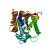
- PDB-2oxm: Crystal structure of a UNG2/modified DNA complex that represent a... -
+
Open data
ID or keywords:
Loading...
-
Basic information
Entry
Database: PDB / ID: 2oxm
Title
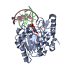
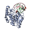
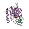
Crystal structure of a UNG2/modified DNA complex that represent a stabilized short-lived extrahelical state in ezymatic DNA base flipping
Components
DNA (5'-D(*AP*AP*AP*GP*AP*TP*(4MF)P*AP*CP*A)-3')
DNA (5'-D(*TP*GP*TP*TP*AP*TP*CP*TP*T)-3')
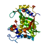
Uracil-DNA glycosylase
Keywords
Hydrolase/DNA / ENZYME DNA COMPLEX / UNG2 / URACIL DNA GLYCOSYLASE / Hydrolase-DNA COMPLEX
Function / homology
Function and homology information
base-excision repair, AP site formation via deaminated base removal / uracil-DNA glycosylase / depyrimidination / Displacement of DNA glycosylase by APEX1 / uracil DNA N-glycosylase activity / single strand break repair / ribosomal small subunit binding / Recognition and association of DNA glycosylase with site containing an affected pyrimidine / Cleavage of the damaged pyrimidine / Chromatin modifications during the maternal to zygotic transition (MZT) ...base-excision repair, AP site formation via deaminated base removal / uracil-DNA glycosylase / depyrimidination / Displacement of DNA glycosylase by APEX1 / uracil DNA N-glycosylase activity / single strand break repair / ribosomal small subunit binding / Recognition and association of DNA glycosylase with site containing an affected pyrimidine / Cleavage of the damaged pyrimidine / Chromatin modifications during the maternal to zygotic transition (MZT) / base-excision repair / damaged DNA binding / negative regulation of apoptotic process / mitochondrion / nucleoplasm / nucleus Similarity search - Function
Uracil-DNA glycosylase family 1 / Uracil DNA glycosylase superfamily / UreE urease accessory protein, C-terminal domain / Uracil-DNA Glycosylase, subunit E / Uracil-DNA glycosylase-like domain / Uracil-DNA glycosylase, active site / Uracil-DNA glycosylase signature. / Uracil-DNA glycosylase-like / Uracil DNA glycosylase superfamily / Uracil-DNA glycosylase-like domain superfamily ...Uracil-DNA glycosylase family 1 / Uracil DNA glycosylase superfamily / UreE urease accessory protein, C-terminal domain / Uracil-DNA Glycosylase, subunit E / Uracil-DNA glycosylase-like domain / Uracil-DNA glycosylase, active site / Uracil-DNA glycosylase signature. / Uracil-DNA glycosylase-like / Uracil DNA glycosylase superfamily / Uracil-DNA glycosylase-like domain superfamily / 3-Layer(aba) Sandwich / Alpha Beta Similarity search - Domain/homology
Mass: 18.015 Da / Num. of mol.: 107 / Source method: isolated from a natural source / Formula: H2O
-
Experimental details
-
Experiment
Experiment
Method: X-RAY DIFFRACTION / Number of used crystals: 1
-
Sample preparation
Crystal
Density Matthews: 2.48 Å3/Da / Density % sol: 50.45 %
Crystal grow
Temperature: 293 K / Method: vapor diffusion, hanging drop / pH: 6.5 Details: A solution of human UNG2 (22 mg/ml) in 50 mM Tris-OAc buffer pH 7.0, 150 mM NaCl and 1mM DTT was mixed with T/M duplex DNA (2.5 mM)m incubate at room Temperature for 30 min. and then ...Details: A solution of human UNG2 (22 mg/ml) in 50 mM Tris-OAc buffer pH 7.0, 150 mM NaCl and 1mM DTT was mixed with T/M duplex DNA (2.5 mM)m incubate at room Temperature for 30 min. and then centrifugate at 10000 g for 5 min.Co-crystallizarion condition were 22% PEG 4000, 100 MM HEPES pH 6.5 and 1mM DTT, VAPOR DIFFUSION, HANGING DROP, temperature 293K
In the structure databanks used in Yorodumi, some data are registered as the other names, "COVID-19 virus" and "2019-nCoV". Here are the details of the virus and the list of structure data.
Jan 31, 2019. EMDB accession codes are about to change! (news from PDBe EMDB page)
EMDB accession codes are about to change! (news from PDBe EMDB page)
The allocation of 4 digits for EMDB accession codes will soon come to an end. Whilst these codes will remain in use, new EMDB accession codes will include an additional digit and will expand incrementally as the available range of codes is exhausted. The current 4-digit format prefixed with “EMD-” (i.e. EMD-XXXX) will advance to a 5-digit format (i.e. EMD-XXXXX), and so on. It is currently estimated that the 4-digit codes will be depleted around Spring 2019, at which point the 5-digit format will come into force.
The EM Navigator/Yorodumi systems omit the EMD- prefix.
Related info.:Q: What is EMD? / ID/Accession-code notation in Yorodumi/EM Navigator
Yorodumi is a browser for structure data from EMDB, PDB, SASBDB, etc.
This page is also the successor to EM Navigator detail page, and also detail information page/front-end page for Omokage search.
The word "yorodu" (or yorozu) is an old Japanese word meaning "ten thousand". "mi" (miru) is to see.
Related info.:EMDB / PDB / SASBDB / Comparison of 3 databanks / Yorodumi Search / Aug 31, 2016. New EM Navigator & Yorodumi / Yorodumi Papers / Jmol/JSmol / Function and homology information / Changes in new EM Navigator and Yorodumi
 Movie
Movie Controller

 Yorodumi
 Open data
 Basic information
 Components
 Keywords
 Function and homology information
 Homo sapiens (human)
 X-RAY DIFFRACTION /
  Authors
 Citation
 Structure visualization
 Downloads & links
 Other downloads


 PDBj
PDBj
 Assembly

 Mass: 18.015 Da / Num. of mol.: 107 / Source method: isolated from a natural source / Formula: H2O
 Sample preparation
 Processing