 Movie
Movie Controller

[English] 日本語
 Yorodumi
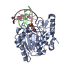
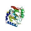
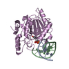
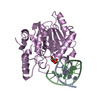
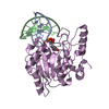
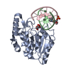
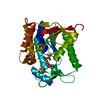
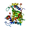
- PDB-1emj: URACIL-DNA GLYCOSYLASE BOUND TO DNA CONTAINING A 4'-THIO-2'DEOXYU... -
+ Open data

- Basic information
| Entry | Database: PDB / ID: 1emj | ||||||
|---|---|---|---|---|---|---|---|
| Title | URACIL-DNA GLYCOSYLASE BOUND TO DNA CONTAINING A 4'-THIO-2'DEOXYURIDINE ANALOG PRODUCT | ||||||
 Components Components |
| ||||||
 Keywords Keywords | hydrolase/DNA / alpha/beta fold / Uracil-DNA Glycosylase / protein/DNA / hydrolase-DNA COMPLEX | ||||||
| Function / homology |  Function and homology information Function and homology informationbase-excision repair, AP site formation via deaminated base removal / uracil-DNA glycosylase / depyrimidination / Displacement of DNA glycosylase by APEX1 / single strand break repair / uracil DNA N-glycosylase activity / isotype switching / ribosomal small subunit binding / somatic hypermutation of immunoglobulin genes / Recognition and association of DNA glycosylase with site containing an affected pyrimidine ...base-excision repair, AP site formation via deaminated base removal / uracil-DNA glycosylase / depyrimidination / Displacement of DNA glycosylase by APEX1 / single strand break repair / uracil DNA N-glycosylase activity / isotype switching / ribosomal small subunit binding / somatic hypermutation of immunoglobulin genes / Recognition and association of DNA glycosylase with site containing an affected pyrimidine / Cleavage of the damaged pyrimidine / Chromatin modifications during the maternal to zygotic transition (MZT) / base-excision repair / damaged DNA binding / negative regulation of apoptotic process / mitochondrion / nucleoplasm / nucleus Similarity search - Function | ||||||
| Biological species |  Homo sapiens (human) Homo sapiens (human) | ||||||
| Method |  X-RAY DIFFRACTION / SYNCHROTRON / Resolution: 2 Å X-RAY DIFFRACTION / SYNCHROTRON / Resolution: 2 Å | ||||||
 Authors Authors | Parikh, S.S. / Walcher, G. / Jones, G.D. / Slupphaug, G. / Krokan, H.E. / Blackburn, G.M. / Tainer, J.A. | ||||||
 Citation Citation | Journal: Proc.Natl.Acad.Sci.USA / Year: 2000 Title: Uracil-DNA glycosylase-DNA substrate and product structures: conformational strain promotes catalytic efficiency by coupled stereoelectronic effects. Authors: Parikh, S.S. / Walcher, G. / Jones, G.D. / Slupphaug, G. / Krokan, H.E. / Blackburn, G.M. / Tainer, J.A. #1: Journal: Embo J. / Year: 1998Title: Base excision repair initiation revealed by crystal structures and binding kinetics of human uracil-DNA glycosylase with DNA Authors: Parikh, S.S. / Mol, C.D. / Slupphaug, G. / Krokan, H.E. / Tainer, J.A. | ||||||
| History |
|
- Structure visualization
| Structure viewer | Molecule: MolmilJmol/JSmol |
|---|
- Downloads & links
-Download
| PDBx/mmCIF format | 1emj.cif.gz | 73.6 KB | Display | PDBx/mmCIF format |
|---|---|---|---|---|
| PDB format | pdb1emj.ent.gz | 51.3 KB | Display | PDB format |
| PDBx/mmJSON format | 1emj.json.gz | Tree view | PDBx/mmJSON format | |
| Others |  Other downloads Other downloads |
-Validation report
| Arichive directory | https://data.pdbj.org/pub/pdb/validation_reports/em/1emjftp://data.pdbj.org/pub/pdb/validation_reports/em/1emj | HTTPS FTP |
|---|
-Related structure data


-Links
 PDBj
PDBj
- Assembly
| Deposited unit | 
| ||||||||||
|---|---|---|---|---|---|---|---|---|---|---|---|
| 1 |
| ||||||||||
| Unit cell |
|
-Components
| #1: DNA chain | Mass: 2619.762 Da / Num. of mol.: 1 / Source method: obtained synthetically |
|---|---|
| #2: DNA chain | Mass: 3070.071 Da / Num. of mol.: 1 / Source method: obtained synthetically |
| #3: Protein | Mass: 25544.137 Da / Num. of mol.: 1 / Mutation: RESIDUES 85-304 Source method: isolated from a genetically manipulated source Details: MITOCHONDRIAL PROTEIN / Source: (gene. exp.) Homo sapiens (human) / Production host:  |
| #4: Chemical | ChemComp-URA /   Mass: 112.087 Da / Num. of mol.: 1 / Source method: obtained synthetically / Formula: C4H4N2O2 Mass: 112.087 Da / Num. of mol.: 1 / Source method: obtained synthetically / Formula: C4H4N2O2 |
| #5: Water | ChemComp-HOH /  Mass: 18.015 Da / Num. of mol.: 161 / Source method: isolated from a natural source / Formula: H2O Mass: 18.015 Da / Num. of mol.: 161 / Source method: isolated from a natural source / Formula: H2O |
-Experimental details
-Experiment
| Experiment | Method: X-RAY DIFFRACTION / Number of used crystals: 1 |
|---|
- Sample preparation
| Crystal | Density Matthews: 2.4 Å3/Da / Density % sol: 48.83 % | |||||||||||||||||||||||||||||||||||
|---|---|---|---|---|---|---|---|---|---|---|---|---|---|---|---|---|---|---|---|---|---|---|---|---|---|---|---|---|---|---|---|---|---|---|---|---|
| Crystal grow | Temperature: 298 K / Method: vapor diffusion, hanging drop / pH: 6.5 Details: PEG 4000, HEPES buffer, NaCl, dioxane, DTT, pH 6.5, VAPOR DIFFUSION, HANGING DROP, temperature 298K | |||||||||||||||||||||||||||||||||||
| Components of the solutions |
| |||||||||||||||||||||||||||||||||||
| Crystal grow | *PLUS Method: vapor diffusion | |||||||||||||||||||||||||||||||||||
| Components of the solutions | *PLUS
|
-Data collection
| Diffraction | Mean temperature: 110 K |
|---|---|
| Diffraction source | Source: SYNCHROTRON / Site: SSRL  / Beamline: BL7-1 / Wavelength: 1.08 / Beamline: BL7-1 / Wavelength: 1.08 |
| Detector | Type: MARRESEARCH / Detector: IMAGE PLATE / Date: Dec 18, 1996 |
| Radiation | Protocol: SINGLE WAVELENGTH / Monochromatic (M) / Laue (L): M / Scattering type: x-ray |
| Radiation wavelength | Wavelength: 1.08 Å / Relative weight: 1 |
| Reflection | Resolution: 2→30 Å / Num. all: 21503 / Num. obs: 21491 / % possible obs: 99.9 % / Observed criterion σ(F): 2 / Observed criterion σ(I): 1 / Redundancy: 6.99 % / Biso Wilson estimate: 19.44 Å2 / Rmerge(I) obs: 0.131 / Net I/σ(I): 6 |
| Reflection shell | Resolution: 2→2.07 Å / Redundancy: 3 % / Rmerge(I) obs: 0.464 / Num. unique all: 2088 / % possible all: 99.8 |
| Reflection | *PLUS Num. measured all: 150204 |
| Reflection shell | *PLUS % possible obs: 99.8 % |
- Processing
| Software |
| |||||||||||||||||||||||||
|---|---|---|---|---|---|---|---|---|---|---|---|---|---|---|---|---|---|---|---|---|---|---|---|---|---|---|
| Refinement | Resolution: 2→20 Å / Cross valid method: THROUGHOUT / σ(F): 2 / Stereochemistry target values: Engh & Huber
| |||||||||||||||||||||||||
| Refinement step | Cycle: LAST / Resolution: 2→20 Å
| |||||||||||||||||||||||||
| Software | *PLUS Name: CNS / Classification: refinement | |||||||||||||||||||||||||
| Refinement | *PLUS Highest resolution: 2 Å / Lowest resolution: 20 Å / σ(F): 2 / % reflection Rfree: 10 % / Rfactor obs: 0.228 | |||||||||||||||||||||||||
| Solvent computation | *PLUS | |||||||||||||||||||||||||
| Displacement parameters | *PLUS | |||||||||||||||||||||||||
| Refine LS restraints | *PLUS
|
