 Movie
Movie Controller

[English] 日本語
 Yorodumi
- PDB-4skn: A NUCLEOTIDE-FLIPPING MECHANISM FROM THE STRUCTURE OF HUMAN URACI... -
+ Open data

- Basic information
| Entry | Database: PDB / ID: 4skn | ||||||
|---|---|---|---|---|---|---|---|
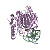
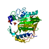
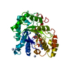
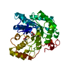
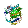
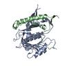
| Title | A NUCLEOTIDE-FLIPPING MECHANISM FROM THE STRUCTURE OF HUMAN URACIL-DNA GLYCOSYLASE BOUND TO DNA | ||||||
 Components Components |
| ||||||
 Keywords Keywords | HYDROLASE/DNA / DNA GLYCOSYLASE / DNA BASE EXCISION REPAIR / URACIL / DNA / HYDROLASE-DNA COMPLEX | ||||||
| Function / homology |  Function and homology information Function and homology informationbase-excision repair, AP site formation via deaminated base removal / uracil-DNA glycosylase / depyrimidination / Displacement of DNA glycosylase by APEX1 / single strand break repair / uracil DNA N-glycosylase activity / isotype switching / ribosomal small subunit binding / somatic hypermutation of immunoglobulin genes / Recognition and association of DNA glycosylase with site containing an affected pyrimidine ...base-excision repair, AP site formation via deaminated base removal / uracil-DNA glycosylase / depyrimidination / Displacement of DNA glycosylase by APEX1 / single strand break repair / uracil DNA N-glycosylase activity / isotype switching / ribosomal small subunit binding / somatic hypermutation of immunoglobulin genes / Recognition and association of DNA glycosylase with site containing an affected pyrimidine / Cleavage of the damaged pyrimidine / Chromatin modifications during the maternal to zygotic transition (MZT) / base-excision repair / damaged DNA binding / negative regulation of apoptotic process / mitochondrion / nucleoplasm / nucleus Similarity search - Function | ||||||
| Biological species |  Homo sapiens (human) Homo sapiens (human) | ||||||
| Method |  X-RAY DIFFRACTION / SYNCHROTRON / MOLECULAR REPLACEMENT / Resolution: 2.9 Å X-RAY DIFFRACTION / SYNCHROTRON / MOLECULAR REPLACEMENT / Resolution: 2.9 Å | ||||||
 Authors Authors | Slupphaug, G. / Mol, C.D. / Kavli, B. / Arvai, A.S. / Krokan, H.E. / Tainer, J.A. | ||||||
 Citation Citation | Journal: Nature / Year: 1996 Title: A nucleotide-flipping mechanism from the structure of human uracil-DNA glycosylase bound to DNA. Authors: Slupphaug, G. / Mol, C.D. / Kavli, B. / Arvai, A.S. / Krokan, H.E. / Tainer, J.A. | ||||||
| History |
|
- Structure visualization
| Structure viewer | Molecule: MolmilJmol/JSmol |
|---|
- Downloads & links
-Download
| PDBx/mmCIF format | 4skn.cif.gz | 73.3 KB | Display | PDBx/mmCIF format |
|---|---|---|---|---|
| PDB format | pdb4skn.ent.gz | 50.5 KB | Display | PDB format |
| PDBx/mmJSON format | 4skn.json.gz | Tree view | PDBx/mmJSON format | |
| Others |  Other downloads Other downloads |
-Validation report
| Arichive directory | https://data.pdbj.org/pub/pdb/validation_reports/sk/4sknftp://data.pdbj.org/pub/pdb/validation_reports/sk/4skn | HTTPS FTP |
|---|
-Related structure data
| Related structure data | 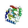 1akzS S: Starting model for refinement |
|---|---|
| Similar structure data |


-Links
 PDBj
PDBj


- Assembly
| Deposited unit | 
| ||||||||||
|---|---|---|---|---|---|---|---|---|---|---|---|
| 1 |
| ||||||||||
| Unit cell |
|
-Components
| #1: DNA chain | Mass: 2998.927 Da / Num. of mol.: 1 / Source method: obtained synthetically |
|---|---|
| #2: DNA chain | Mass: 2998.984 Da / Num. of mol.: 1 / Source method: obtained synthetically |
| #3: Protein | Mass: 25587.188 Da / Num. of mol.: 1 / Mutation: P82M, W83E, G84F, D145N, L272R Source method: isolated from a genetically manipulated source Source: (gene. exp.) Homo sapiens (human) / Production host:  |
| #4: Chemical | ChemComp-URA /   Mass: 112.087 Da / Num. of mol.: 1 / Source method: obtained synthetically / Formula: C4H4N2O2 Mass: 112.087 Da / Num. of mol.: 1 / Source method: obtained synthetically / Formula: C4H4N2O2 |
| #5: Water | ChemComp-HOH /  Mass: 18.015 Da / Num. of mol.: 75 / Source method: isolated from a natural source / Formula: H2O Mass: 18.015 Da / Num. of mol.: 75 / Source method: isolated from a natural source / Formula: H2O |
-Experimental details
-Experiment
| Experiment | Method: X-RAY DIFFRACTION / Number of used crystals: 1 |
|---|
- Sample preparation
| Crystal | Density Matthews: 2.44 Å3/Da / Density % sol: 49.68 % | ||||||||||||||||||||
|---|---|---|---|---|---|---|---|---|---|---|---|---|---|---|---|---|---|---|---|---|---|
| Crystal grow | pH: 6.5 / Details: pH 6.5 | ||||||||||||||||||||
| Crystal grow | *PLUS Temperature: 22 ℃ / Method: vapor diffusion, sitting drop | ||||||||||||||||||||
| Components of the solutions | *PLUS
|
-Data collection
| Diffraction | Mean temperature: 100 K |
|---|---|
| Diffraction source | Source: SYNCHROTRON / Site: SSRL  / Beamline: BL7-1 / Wavelength: 1.08 / Beamline: BL7-1 / Wavelength: 1.08 |
| Detector | Date: Apr 15, 1996 |
| Radiation | Protocol: SINGLE WAVELENGTH / Monochromatic (M) / Laue (L): M / Scattering type: x-ray |
| Radiation wavelength | Wavelength: 1.08 Å / Relative weight: 1 |
| Reflection | Resolution: 2.9→40 Å / Num. obs: 7140 / % possible obs: 97 % / Redundancy: 3.1 % / Rsym value: 0.108 / Net I/σ(I): 7.9 |
| Reflection shell | Resolution: 2.9→3 Å / Rsym value: 0.398 / % possible all: 98 |
| Reflection | *PLUS Num. measured all: 21922 / Rmerge(I) obs: 0.108 |
| Reflection shell | *PLUS % possible obs: 98 % / Rmerge(I) obs: 0.398 |
- Processing
| Software |
| ||||||||||||||||||||||||||||||||||||||||||||||||||||||||||||
|---|---|---|---|---|---|---|---|---|---|---|---|---|---|---|---|---|---|---|---|---|---|---|---|---|---|---|---|---|---|---|---|---|---|---|---|---|---|---|---|---|---|---|---|---|---|---|---|---|---|---|---|---|---|---|---|---|---|---|---|---|---|
| Refinement | Method to determine structure: MOLECULAR REPLACEMENT Starting model: PDB ENTRY 1AKZ Resolution: 2.9→20 Å / Data cutoff high absF: 100000 / Data cutoff low absF: 0.1 / Cross valid method: THROUGHOUT / σ(F): 2.9 Details: THE ELECTRON DENSITY FOR THE DNA 5' OF THE URACIL-CONTAINING NUCLEOTIDE IS POOR. THE COBALAMIN HAS BEEN MODELLED AS THE 5-COORDINATES REDUCED COB(II)ALAMIN (B12R), SINCE THERE IS NO ELECTRON ...Details: THE ELECTRON DENSITY FOR THE DNA 5' OF THE URACIL-CONTAINING NUCLEOTIDE IS POOR. THE COBALAMIN HAS BEEN MODELLED AS THE 5-COORDINATES REDUCED COB(II)ALAMIN (B12R), SINCE THERE IS NO ELECTRON DENSITY FOR AN ADENOSYL GROUP LINKED TO THE COBALT ATOM. HOWEVER, A HALF-OCCUPIED 5'-DEOXYADENOSINE HAS BEEN BUILT INTO ELECTRON DENSITY IN THE ACTIVE SITE (RESIDUES A803 AND AND C803). RESIDUES A801 AND A802 (AND C801, C802) REPRESENT AN EQUIMOLAR MIXTURE OF SUBSTRATE AND PRODUCT, SUCCINYL- AND METHYLMALONYL-COENZYME A.
| ||||||||||||||||||||||||||||||||||||||||||||||||||||||||||||
| Displacement parameters |
| ||||||||||||||||||||||||||||||||||||||||||||||||||||||||||||
| Refinement step | Cycle: LAST / Resolution: 2.9→20 Å
| ||||||||||||||||||||||||||||||||||||||||||||||||||||||||||||
| Refine LS restraints |
| ||||||||||||||||||||||||||||||||||||||||||||||||||||||||||||
| LS refinement shell | Resolution: 2.9→3.03 Å / Total num. of bins used: 8
| ||||||||||||||||||||||||||||||||||||||||||||||||||||||||||||
| Xplor file |
| ||||||||||||||||||||||||||||||||||||||||||||||||||||||||||||
| Software | *PLUS Name: X-PLOR / Version: 3.851 / Classification: refinement | ||||||||||||||||||||||||||||||||||||||||||||||||||||||||||||
| Refine LS restraints | *PLUS
|
