 Movie
Movie Controller

[English] 日本語
 Yorodumi
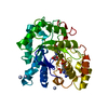
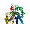
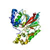
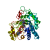
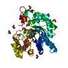
- PDB-6kbl: Structure-function study of AKR4C14, an aldo-keto reductase from ... -
+ Open data

- Basic information
| Entry | Database: PDB / ID: 6kbl | ||||||
|---|---|---|---|---|---|---|---|
| Title | Structure-function study of AKR4C14, an aldo-keto reductase from Thai Jasmine rice (Oryza sativa L. ssp. Indica cv. KDML105) | ||||||
 Components Components | Aldo-keto reductase | ||||||
 Keywords Keywords | OXIDOREDUCTASE / Aldo-keto reductase (AKR) / Aldehyde reductase / Aldose reductase / rice AKR / AKR4C14 | ||||||
| Function / homology |  Function and homology information Function and homology information | ||||||
| Biological species |  | ||||||
| Method |  X-RAY DIFFRACTION / SYNCHROTRON / MOLECULAR REPLACEMENT / Resolution: 1.7 Å X-RAY DIFFRACTION / SYNCHROTRON / MOLECULAR REPLACEMENT / Resolution: 1.7 Å | ||||||
 Authors Authors | Songsiriritthigul, C. / Narawongsanont, R. / Guan, H.H. / Chen, C.J. | ||||||
 Citation Citation | Journal: Acta Crystallogr D Struct Biol / Year: 2020 Title: Structure-function study of AKR4C14, an aldo-keto reductase from Thai jasmine rice (Oryza sativa L. ssp. indica cv. KDML105). Authors: Songsiriritthigul, C. / Narawongsanont, R. / Tantitadapitak, C. / Guan, H.H. / Chen, C.J. #1: Journal: Protein J. / Year: 2017Title: Characterization of AKR4C15, a Novel Member of Aldo-Keto Reductase, in Comparison with Other Rice AKR(s). Authors: Auiyawong, B. / Narawongsanont, R. / Tantitadapitak, C. | ||||||
| History |
|
- Structure visualization
| Structure viewer | Molecule: MolmilJmol/JSmol |
|---|
- Downloads & links
-Download
| PDBx/mmCIF format | 6kbl.cif.gz | 80.7 KB | Display | PDBx/mmCIF format |
|---|---|---|---|---|
| PDB format | pdb6kbl.ent.gz | 58.1 KB | Display | PDB format |
| PDBx/mmJSON format | 6kbl.json.gz | Tree view | PDBx/mmJSON format | |
| Others |  Other downloads Other downloads |
-Validation report
| Arichive directory | https://data.pdbj.org/pub/pdb/validation_reports/kb/6kblftp://data.pdbj.org/pub/pdb/validation_reports/kb/6kbl | HTTPS FTP |
|---|
-Related structure data
| Related structure data |  3h7uS S: Starting model for refinement |
|---|---|
| Similar structure data |


-Links
 PDBj
PDBj


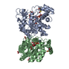
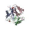
- Assembly
| Deposited unit | 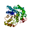
| ||||||||
|---|---|---|---|---|---|---|---|---|---|
| 1 |
| ||||||||
| Unit cell |
|
-Components
| #1: Protein | Mass: 34725.684 Da / Num. of mol.: 1 Source method: isolated from a genetically manipulated source Source: (gene. exp.) Gene: AKR2, OsI_04428 / Plasmid: PET28B(+) / Production host:  |
|---|---|
| #2: Chemical | ChemComp-CAC /   Mass: 136.989 Da / Num. of mol.: 1 / Source method: obtained synthetically / Formula: C2H6AsO2 Mass: 136.989 Da / Num. of mol.: 1 / Source method: obtained synthetically / Formula: C2H6AsO2 |
| #3: Chemical | ChemComp-ACT /   Mass: 59.044 Da / Num. of mol.: 1 / Source method: obtained synthetically / Formula: C2H3O2 Mass: 59.044 Da / Num. of mol.: 1 / Source method: obtained synthetically / Formula: C2H3O2 |
| #4: Chemical | ChemComp-GOL /   Mass: 92.094 Da / Num. of mol.: 1 / Source method: obtained synthetically / Formula: C3H8O3 Mass: 92.094 Da / Num. of mol.: 1 / Source method: obtained synthetically / Formula: C3H8O3 |
| #5: Water | ChemComp-HOH /  Mass: 18.015 Da / Num. of mol.: 204 / Source method: isolated from a natural source / Formula: H2O Mass: 18.015 Da / Num. of mol.: 204 / Source method: isolated from a natural source / Formula: H2O |
| Has ligand of interest | N |
| Sequence details | The amino acid residue translated from gene is Glu74. But the amino acid that be properly rebuilt ...The amino acid residue translated from gene is Glu74. But the amino acid that be properly rebuilt in the map is Ala74 (appeared in the PDB Coordinates), owing to the disorder of its side chain. |
-Experimental details
-Experiment
| Experiment | Method: X-RAY DIFFRACTION / Number of used crystals: 1 |
|---|
- Sample preparation
| Crystal | Density Matthews: 2.12 Å3/Da / Density % sol: 42.1 % / Description: Rectangular morphology |
|---|---|
| Crystal grow | Temperature: 277 K / Method: batch mode / pH: 6.5 Details: 0.2 M sodium acetate, 0.1 M sodium cacodylate pH 6.5, 30%(w/v) PEG 8000 |
-Data collection
| Diffraction | Mean temperature: 110 K / Ambient temp details: Nitrogen stream / Serial crystal experiment: N |
|---|---|
| Diffraction source | Source: SYNCHROTRON / Site: NSRRC  / Beamline: BL13B1 / Wavelength: 1 Å / Beamline: BL13B1 / Wavelength: 1 Å |
| Detector | Type: ADSC QUANTUM 315 / Detector: CCD / Date: Jul 21, 2016 |
| Radiation | Monochromator: DCM / Protocol: SINGLE WAVELENGTH / Monochromatic (M) / Laue (L): M / Scattering type: x-ray |
| Radiation wavelength | Wavelength: 1 Å / Relative weight: 1 |
| Reflection | Resolution: 1.7→30 Å / Num. obs: 33541 / % possible obs: 99.1 % / Redundancy: 5.6 % / Rmerge(I) obs: 0.275 / Net I/σ(I): 18.28 |
| Reflection shell | Resolution: 1.7→1.73 Å / Redundancy: 4.9 % / Rmerge(I) obs: 0.073 / Mean I/σ(I) obs: 4.53 / Num. unique obs: 1639 / % possible all: 98.2 |
- Processing
| Software |
| ||||||||||||||||||||
|---|---|---|---|---|---|---|---|---|---|---|---|---|---|---|---|---|---|---|---|---|---|
| Refinement | Method to determine structure: MOLECULAR REPLACEMENT Starting model: 3H7U Resolution: 1.7→29.24 Å / Cross valid method: THROUGHOUT
| ||||||||||||||||||||
| Displacement parameters | Biso mean: 20.936 Å2 | ||||||||||||||||||||
| Refinement step | Cycle: LAST / Resolution: 1.7→29.24 Å
| ||||||||||||||||||||
| LS refinement shell | Resolution: 1.7→1.74 Å
|
