 Movie
Movie Controller

[English] 日本語
 Yorodumi
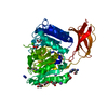
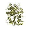
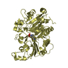
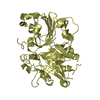
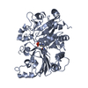
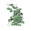
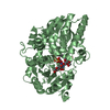
- PDB-1q32: Crystal Structure Analysis of the Yeast Tyrosyl-DNA Phosphodiesterase -
+ Open data

- Basic information
| Entry | Database: PDB / ID: 1q32 | ||||||
|---|---|---|---|---|---|---|---|
| Title | Crystal Structure Analysis of the Yeast Tyrosyl-DNA Phosphodiesterase | ||||||
 Components Components | tyrosyl-DNA phosphodiesterase | ||||||
 Keywords Keywords | REPLICATION / TRANSCRIPTION / HYDROLASE / Tyrosyl-DNA Phosphodiesterase / TDP / DNA repair | ||||||
| Function / homology |  Function and homology information Function and homology information5'-tyrosyl-DNA phosphodiesterase activity / 3'-tyrosyl-DNA phosphodiesterase activity / Hydrolases; Acting on ester bonds; Phosphoric-diester hydrolases / exonuclease activity / single-stranded DNA binding / double-stranded DNA binding / DNA repair / mitochondrion / nucleus Similarity search - Function | ||||||
| Biological species |  | ||||||
| Method |  X-RAY DIFFRACTION / SYNCHROTRON / MAD / Resolution: 2.03 Å X-RAY DIFFRACTION / SYNCHROTRON / MAD / Resolution: 2.03 Å | ||||||
 Authors Authors | He, X. / Babaoglu, K. / Price, A. / Nitiss, K.C. / Nitiss, J.L. / White, S.W. | ||||||
 Citation Citation | Journal: J.Mol.Biol. / Year: 2007 Title: Mutation of a conserved active site residue converts tyrosyl-DNA phosphodiesterase I into a DNA topoisomerase I-dependent poison Authors: He, X. / van Waardenburg, R.C. / Babaoglu, K. / Price, A.C. / Nitiss, K.C. / Nitiss, J.L. / Bjornsti, M.A. / White, S.W. | ||||||
| History |
|
- Structure visualization
| Structure viewer | Molecule: MolmilJmol/JSmol |
|---|
- Downloads & links
-Download
| PDBx/mmCIF format | 1q32.cif.gz | 363.7 KB | Display | PDBx/mmCIF format |
|---|---|---|---|---|
| PDB format | pdb1q32.ent.gz | 293.3 KB | Display | PDB format |
| PDBx/mmJSON format | 1q32.json.gz | Tree view | PDBx/mmJSON format | |
| Others |  Other downloads Other downloads |
-Validation report
| Summary document | 1q32_validation.pdf.gz | 450.8 KB | Display | wwPDB validaton report |
|---|---|---|---|---|
| Full document | 1q32_full_validation.pdf.gz | 480.5 KB | Display | |
| Data in XML | 1q32_validation.xml.gz | 69.1 KB | Display | |
| Data in CIF | 1q32_validation.cif.gz | 97.4 KB | Display | |
| Arichive directory | https://data.pdbj.org/pub/pdb/validation_reports/q3/1q32ftp://data.pdbj.org/pub/pdb/validation_reports/q3/1q32 | HTTPS FTP |
-Related structure data
| Similar structure data |
|---|


-Links
 PDBj
PDBj- Assembly
| Deposited unit | 
| ||||||||||
|---|---|---|---|---|---|---|---|---|---|---|---|
| 1 | 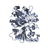
| ||||||||||
| 2 | 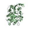
| ||||||||||
| 3 | 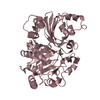
| ||||||||||
| 4 | 
| ||||||||||
| Unit cell |
| ||||||||||
| Details | There are 4 biological assemblies in the asymmetric unit. Each is a monomer, labeled A, B, C, or D. |
-Components
| #1: Protein | Mass: 62414.082 Da / Num. of mol.: 4 Source method: isolated from a genetically manipulated source Source: (gene. exp.) Gene: TDP1 / Plasmid: pET-23b (Novagen) / Production host:  References: UniProt: P38319, Hydrolases; Acting on ester bonds; Phosphoric-diester hydrolases #2: Water | ChemComp-HOH / |  Mass: 18.015 Da / Num. of mol.: 862 / Source method: isolated from a natural source / Formula: H2O Mass: 18.015 Da / Num. of mol.: 862 / Source method: isolated from a natural source / Formula: H2O |
|---|
-Experimental details
-Experiment
| Experiment | Method: X-RAY DIFFRACTION / Number of used crystals: 2 |
|---|
- Sample preparation
| Crystal | Density Matthews: 2.35 Å3/Da / Density % sol: 47.2 % |
|---|---|
| Crystal grow | Temperature: 291.15 K / Method: evaporation / pH: 7.8 Details: PEG 4000, Hepes, LiSO4, DTT, 1.6.Hexanediol , pH 7.8, EVAPORATION, temperature 291.15K |
-Data collection
| Diffraction |
| ||||||||||||||||||
|---|---|---|---|---|---|---|---|---|---|---|---|---|---|---|---|---|---|---|---|
| Diffraction source |
| ||||||||||||||||||
| Detector |
| ||||||||||||||||||
| Radiation |
| ||||||||||||||||||
| Radiation wavelength |
| ||||||||||||||||||
| Reflection | Resolution: 1.8→50 Å / Num. all: 155815 / Num. obs: 155815 / % possible obs: 90.4 % / Observed criterion σ(I): -3 / Redundancy: 3.4 % / Rmerge(I) obs: 0.067 | ||||||||||||||||||
| Reflection shell | Resolution: 1.8→1.86 Å / % possible all: 54.3 |

- Processing
| Software |
| ||||||||||||||||||||||||||||||||||||
|---|---|---|---|---|---|---|---|---|---|---|---|---|---|---|---|---|---|---|---|---|---|---|---|---|---|---|---|---|---|---|---|---|---|---|---|---|---|
| Refinement | Method to determine structure: MAD / Resolution: 2.03→30 Å / Cross valid method: THROUGHOUT / σ(F): 0 / σ(I): 0 / Stereochemistry target values: Engh & Huber
| ||||||||||||||||||||||||||||||||||||
| Refinement step | Cycle: LAST / Resolution: 2.03→30 Å
| ||||||||||||||||||||||||||||||||||||
| Refine LS restraints |
|
