 Movie
Movie Controller

[English] 日本語
 Yorodumi
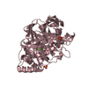
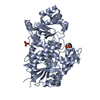
- PDB-5v2j: Crystal structure of UDP-glucosyltransferase, UGT74F2 (T15S), wit... -
+ Open data

- Basic information
| Entry | Database: PDB / ID: 5v2j | |||||||||
|---|---|---|---|---|---|---|---|---|---|---|
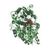
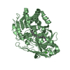
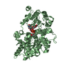
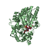
| Title | Crystal structure of UDP-glucosyltransferase, UGT74F2 (T15S), with UDP and 2-bromobenzoic acid | |||||||||
 Components Components | UDP-glycosyltransferase 74F2 | |||||||||
 Keywords Keywords | TRANSFERASE / UDP-glucosyltransferase / Salicylic acid / Salicylic acid glucoside / Salicylic acid glucose ester | |||||||||
| Function / homology |  Function and homology information Function and homology information: / salicylic acid glucosyltransferase (ester-forming) activity / salicylic acid glucosyltransferase (glucoside-forming) activity / benzoic acid glucosyltransferase activity / UDP-glucose:4-aminobenzoate acylglucosyltransferase activity / nicotinate-O-glucosyltransferase activity / benzoate metabolic process / positive regulation of seed germination / quercetin 3-O-glucosyltransferase activity / quercetin 7-O-glucosyltransferase activity ...: / salicylic acid glucosyltransferase (ester-forming) activity / salicylic acid glucosyltransferase (glucoside-forming) activity / benzoic acid glucosyltransferase activity / UDP-glucose:4-aminobenzoate acylglucosyltransferase activity / nicotinate-O-glucosyltransferase activity / benzoate metabolic process / positive regulation of seed germination / quercetin 3-O-glucosyltransferase activity / quercetin 7-O-glucosyltransferase activity / salicylic acid metabolic process / UDP-glucosyltransferase activity / Transferases; Glycosyltransferases; Hexosyltransferases / cytosol / cytoplasm Similarity search - Function | |||||||||
| Biological species |  | |||||||||
| Method |  X-RAY DIFFRACTION / SYNCHROTRON / MOLECULAR REPLACEMENT / Resolution: 1.8 Å X-RAY DIFFRACTION / SYNCHROTRON / MOLECULAR REPLACEMENT / Resolution: 1.8 Å | |||||||||
 Authors Authors | George Thompson, A.M. / Iancu, C.V. / Dean, J.V. / Choe, J. | |||||||||
 Citation Citation | Journal: Sci Rep / Year: 2017 Title: Differences in salicylic acid glucose conjugations by UGT74F1 and UGT74F2 from Arabidopsis thaliana. Authors: George Thompson, A.M. / Iancu, C.V. / Neet, K.E. / Dean, J.V. / Choe, J.Y. | |||||||||
| History |
|
- Structure visualization
| Structure viewer | Molecule: MolmilJmol/JSmol |
|---|
- Downloads & links
-Download
| PDBx/mmCIF format | 5v2j.cif.gz | 377.8 KB | Display | PDBx/mmCIF format |
|---|---|---|---|---|
| PDB format | pdb5v2j.ent.gz | 305.4 KB | Display | PDB format |
| PDBx/mmJSON format | 5v2j.json.gz | Tree view | PDBx/mmJSON format | |
| Others |  Other downloads Other downloads |
-Validation report
| Arichive directory | https://data.pdbj.org/pub/pdb/validation_reports/v2/5v2jftp://data.pdbj.org/pub/pdb/validation_reports/v2/5v2j | HTTPS FTP |
|---|
-Related structure data
| Related structure data | 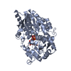 5u6mC 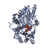 5u6nC  5u6sSC 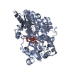 5v2kC C: citing same article ( S: Starting model for refinement |
|---|---|
| Similar structure data |


-Links
 PDBj
PDBj


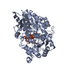
- Assembly
| Deposited unit | 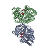
| ||||||||
|---|---|---|---|---|---|---|---|---|---|
| 1 | 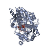
| ||||||||
| 2 | 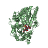
| ||||||||
| Unit cell |
|
-Components
-Protein , 1 types, 2 molecules AB
| #1: Protein | Mass: 50807.594 Da / Num. of mol.: 2 / Mutation: T15S Source method: isolated from a genetically manipulated source Source: (gene. exp.)  References: UniProt: O22822, Transferases; Glycosyltransferases; Hexosyltransferases |
|---|
-Sugars , 2 types, 4 molecules 
| #2: Polysaccharide | 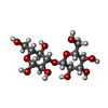  Source method: isolated from a genetically manipulated source Details: oligosaccharide / References: beta-laminaribiose #3: Sugar |  Type: D-saccharide, beta linking / Mass: 180.156 Da / Num. of mol.: 2 / Source method: obtained synthetically / Formula: C6H12O6 Type: D-saccharide, beta linking / Mass: 180.156 Da / Num. of mol.: 2 / Source method: obtained synthetically / Formula: C6H12O6 |
|---|
-Non-polymers , 3 types, 175 molecules 
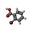

| #4: Chemical |  Type: RNA linking / Mass: 404.161 Da / Num. of mol.: 2 / Source method: isolated from a natural source / Formula: C9H14N2O12P2 / Comment: UDP*YM Type: RNA linking / Mass: 404.161 Da / Num. of mol.: 2 / Source method: isolated from a natural source / Formula: C9H14N2O12P2 / Comment: UDP*YM#5: Chemical |  Mass: 201.017 Da / Num. of mol.: 2 / Source method: obtained synthetically / Formula: C7H5BrO2 Mass: 201.017 Da / Num. of mol.: 2 / Source method: obtained synthetically / Formula: C7H5BrO2#6: Water | ChemComp-HOH / | Mass: 18.015 Da / Num. of mol.: 171 / Source method: isolated from a natural source / Formula: H2O |
|---|
-Details
| Has protein modification | Y |
|---|
-Experimental details
-Experiment
| Experiment | Method: X-RAY DIFFRACTION / Number of used crystals: 1 |
|---|
- Sample preparation
| Crystal | Density Matthews: 2.28 Å3/Da / Density % sol: 46.14 % |
|---|---|
| Crystal grow | Temperature: 293 K / Method: vapor diffusion, hanging drop / pH: 5.5 Details: 22-26 %(w/v) PEG3350, 0.2 M ammonium acetate, 0.1 M MES |
-Data collection
| Diffraction | Mean temperature: 100 K |
|---|---|
| Diffraction source | Source: SYNCHROTRON / Site: APS  / Beamline: 23-ID-B / Wavelength: 0.99187 Å / Beamline: 23-ID-B / Wavelength: 0.99187 Å |
| Detector | Type: DECTRIS EIGER X 16M / Detector: PIXEL / Date: Feb 5, 2017 |
| Radiation | Protocol: SINGLE WAVELENGTH / Monochromatic (M) / Laue (L): M / Scattering type: x-ray |
| Radiation wavelength | Wavelength: 0.99187 Å / Relative weight: 1 |
| Reflection | Resolution: 1.8→50 Å / Num. obs: 90391 / % possible obs: 99.4 % / Redundancy: 5.2 % / Net I/σ(I): 19.2 |
- Processing
| Software |
| |||||||||||||||||||||||||||||||||||||||||||||||||||||||||||||||||||||||||||||||||||||||||||||||||||||||||||||||||||||||||||||||||||||||||||||||||||||||||||||||||||||||||||||||||||||||||||||||||||||||||||||||||||||||||
|---|---|---|---|---|---|---|---|---|---|---|---|---|---|---|---|---|---|---|---|---|---|---|---|---|---|---|---|---|---|---|---|---|---|---|---|---|---|---|---|---|---|---|---|---|---|---|---|---|---|---|---|---|---|---|---|---|---|---|---|---|---|---|---|---|---|---|---|---|---|---|---|---|---|---|---|---|---|---|---|---|---|---|---|---|---|---|---|---|---|---|---|---|---|---|---|---|---|---|---|---|---|---|---|---|---|---|---|---|---|---|---|---|---|---|---|---|---|---|---|---|---|---|---|---|---|---|---|---|---|---|---|---|---|---|---|---|---|---|---|---|---|---|---|---|---|---|---|---|---|---|---|---|---|---|---|---|---|---|---|---|---|---|---|---|---|---|---|---|---|---|---|---|---|---|---|---|---|---|---|---|---|---|---|---|---|---|---|---|---|---|---|---|---|---|---|---|---|---|---|---|---|---|---|---|---|---|---|---|---|---|---|---|---|---|---|---|---|---|
| Refinement | Method to determine structure: MOLECULAR REPLACEMENT Starting model: 5U6S Resolution: 1.8→42.211 Å / SU ML: 0.28 / Cross valid method: FREE R-VALUE / σ(F): 1.35 / Phase error: 27.22 / Stereochemistry target values: ML
| |||||||||||||||||||||||||||||||||||||||||||||||||||||||||||||||||||||||||||||||||||||||||||||||||||||||||||||||||||||||||||||||||||||||||||||||||||||||||||||||||||||||||||||||||||||||||||||||||||||||||||||||||||||||||
| Solvent computation | Shrinkage radii: 0.9 Å / VDW probe radii: 1.11 Å / Solvent model: FLAT BULK SOLVENT MODEL | |||||||||||||||||||||||||||||||||||||||||||||||||||||||||||||||||||||||||||||||||||||||||||||||||||||||||||||||||||||||||||||||||||||||||||||||||||||||||||||||||||||||||||||||||||||||||||||||||||||||||||||||||||||||||
| Refinement step | Cycle: LAST / Resolution: 1.8→42.211 Å
| |||||||||||||||||||||||||||||||||||||||||||||||||||||||||||||||||||||||||||||||||||||||||||||||||||||||||||||||||||||||||||||||||||||||||||||||||||||||||||||||||||||||||||||||||||||||||||||||||||||||||||||||||||||||||
| Refine LS restraints |
| |||||||||||||||||||||||||||||||||||||||||||||||||||||||||||||||||||||||||||||||||||||||||||||||||||||||||||||||||||||||||||||||||||||||||||||||||||||||||||||||||||||||||||||||||||||||||||||||||||||||||||||||||||||||||
| LS refinement shell |
| |||||||||||||||||||||||||||||||||||||||||||||||||||||||||||||||||||||||||||||||||||||||||||||||||||||||||||||||||||||||||||||||||||||||||||||||||||||||||||||||||||||||||||||||||||||||||||||||||||||||||||||||||||||||||
| Refinement TLS params. | Method: refined / Origin x: -10.2914 Å / Origin y: -8.1544 Å / Origin z: 17.9883 Å
| |||||||||||||||||||||||||||||||||||||||||||||||||||||||||||||||||||||||||||||||||||||||||||||||||||||||||||||||||||||||||||||||||||||||||||||||||||||||||||||||||||||||||||||||||||||||||||||||||||||||||||||||||||||||||
| Refinement TLS group | Selection details: all |
