 Movie
Movie Controller

[English] 日本語
 Yorodumi
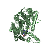
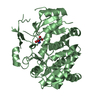
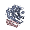
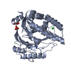
- PDB-1pzm: Crystal structure of HGPRT-ase from Leishmania tarentolae in comp... -
+ Open data

- Basic information
| Entry | Database: PDB / ID: 1pzm | ||||||
|---|---|---|---|---|---|---|---|
| Title | Crystal structure of HGPRT-ase from Leishmania tarentolae in complex with GMP | ||||||
 Components Components | hypoxanthine-guanine phosphoribosyltransferase | ||||||
 Keywords Keywords | TRANSFERASE | ||||||
| Function / homology |  Function and homology information Function and homology informationhypoxanthine phosphoribosyltransferase / guanine salvage / hypoxanthine metabolic process / hypoxanthine phosphoribosyltransferase activity / GMP salvage / IMP salvage / purine ribonucleoside salvage / nucleotide binding / magnesium ion binding / cytosol Similarity search - Function | ||||||
| Biological species |  Leishmania tarentolae (eukaryote) Leishmania tarentolae (eukaryote) | ||||||
| Method |  X-RAY DIFFRACTION / SYNCHROTRON / MOLECULAR REPLACEMENT / Resolution: 2.1 Å X-RAY DIFFRACTION / SYNCHROTRON / MOLECULAR REPLACEMENT / Resolution: 2.1 Å | ||||||
 Authors Authors | Monzani, P.S. / Trapani, S. / Oliva, G. / Thiemann, O.H. | ||||||
 Citation Citation | Journal: Bmc Struct.Biol. / Year: 2007 Title: Crystal structure of Leishmania tarentolae hypoxanthine-guanine phosphoribosyltransferase. Authors: Monzani, P.S. / Trapani, S. / Thiemann, O.H. / Oliva, G. #1: Journal: BIOCHIM.BIOPHYS.ACTA / Year: 2002Title: Cloning, characterization and preliminary crystallographic analysis of Leishmania hypoxanthine-guanine phosphoribosyltransferase Authors: Monzani, P.S. / Alfonzo, J.D. / Simpson, L. / Oliva, G. / Thiemann, O.H. | ||||||
| History |
|
- Structure visualization
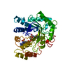
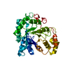
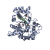
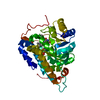
| Structure viewer | Molecule: MolmilJmol/JSmol |
|---|
- Downloads & links
-Download
| PDBx/mmCIF format | 1pzm.cif.gz | 152.8 KB | Display | PDBx/mmCIF format |
|---|---|---|---|---|
| PDB format | pdb1pzm.ent.gz | 120.4 KB | Display | PDB format |
| PDBx/mmJSON format | 1pzm.json.gz | Tree view | PDBx/mmJSON format | |
| Others |  Other downloads Other downloads |
-Validation report
| Summary document | 1pzm_validation.pdf.gz | 1.2 MB | Display | wwPDB validaton report |
|---|---|---|---|---|
| Full document | 1pzm_full_validation.pdf.gz | 1.3 MB | Display | |
| Data in XML | 1pzm_validation.xml.gz | 17.9 KB | Display | |
| Data in CIF | 1pzm_validation.cif.gz | 25.8 KB | Display | |
| Arichive directory | https://data.pdbj.org/pub/pdb/validation_reports/pz/1pzmftp://data.pdbj.org/pub/pdb/validation_reports/pz/1pzm | HTTPS FTP |
-Related structure data
| Related structure data | 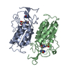 1tc1S S: Starting model for refinement |
|---|---|
| Similar structure data |
-Links
 PDBj
PDBj

- Assembly
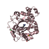
| Deposited unit | 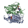
| ||||||||
|---|---|---|---|---|---|---|---|---|---|
| 1 |
| ||||||||
| Unit cell |
| ||||||||
| Details | The asymmetric unit content represents the biological assembly, which is a dimer. |
-Components
| #1: Protein | Mass: 23660.475 Da / Num. of mol.: 2 Source method: isolated from a genetically manipulated source Source: (gene. exp.) Leishmania tarentolae (eukaryote) / Gene: hgprt / Plasmid: pET29a(+) / Species (production host): Escherichia coli / Production host:  References: UniProt: Q9NJI5, hypoxanthine phosphoribosyltransferase #2: Chemical |   Mass: 363.221 Da / Num. of mol.: 2 / Source method: obtained synthetically / Formula: C10H14N5O8P Mass: 363.221 Da / Num. of mol.: 2 / Source method: obtained synthetically / Formula: C10H14N5O8P#3: Water | ChemComp-HOH / |  Mass: 18.015 Da / Num. of mol.: 243 / Source method: isolated from a natural source / Formula: H2O Mass: 18.015 Da / Num. of mol.: 243 / Source method: isolated from a natural source / Formula: H2O |
|---|
-Experimental details
-Experiment
| Experiment | Method: X-RAY DIFFRACTION / Number of used crystals: 1 |
|---|
- Sample preparation
| Crystal | Density Matthews: 2.3 Å3/Da / Density % sol: 46.45 % |
|---|---|
| Crystal grow | Temperature: 291 K / Method: vapor diffusion, hanging drop / pH: 5.6 Details: PEG 4000, i-propanol, glycerol, pH 5.6, VAPOR DIFFUSION, HANGING DROP, temperature 291K |
-Data collection
| Diffraction | Mean temperature: 100 K |
|---|---|
| Diffraction source | Source: SYNCHROTRON / Site: LNLS  / Beamline: D03B-MX1 / Wavelength: 1.537 Å / Beamline: D03B-MX1 / Wavelength: 1.537 Å |
| Detector | Type: MARRESEARCH / Detector: IMAGE PLATE / Date: Aug 16, 2001 |
| Radiation | Monochromator: Si 111 CHANNEL / Protocol: SINGLE WAVELENGTH / Monochromatic (M) / Laue (L): M / Scattering type: x-ray |
| Radiation wavelength | Wavelength: 1.537 Å / Relative weight: 1 |
| Reflection | Resolution: 2.1→48.4 Å / Num. all: 24801 / Num. obs: 24231 / % possible obs: 92.8 % / Observed criterion σ(F): 1 |
| Reflection shell | Resolution: 2.1→2.16 Å / Redundancy: 5.5 % / % possible all: 96.8 |
- Processing
| Software |
| |||||||||||||||||||||||||||||||||||||||||||||||||||||||||||||||||||||||||||
|---|---|---|---|---|---|---|---|---|---|---|---|---|---|---|---|---|---|---|---|---|---|---|---|---|---|---|---|---|---|---|---|---|---|---|---|---|---|---|---|---|---|---|---|---|---|---|---|---|---|---|---|---|---|---|---|---|---|---|---|---|---|---|---|---|---|---|---|---|---|---|---|---|---|---|---|---|
| Refinement | Method to determine structure: MOLECULAR REPLACEMENT Starting model: PDB ENTRY 1TC1 Resolution: 2.1→48.22 Å / Cor.coef. Fo:Fc: 0.938 / Cor.coef. Fo:Fc free: 0.92 / SU B: 4.105 / SU ML: 0.109 / Cross valid method: THROUGHOUT / σ(F): 0 / ESU R: 0.189 / ESU R Free: 0.166 / Stereochemistry target values: MAXIMUM LIKELIHOOD / Details: HYDROGENS HAVE BEEN ADDED IN THE RIDING POSITIONS
| |||||||||||||||||||||||||||||||||||||||||||||||||||||||||||||||||||||||||||
| Solvent computation | Ion probe radii: 0.8 Å / Shrinkage radii: 0.8 Å / VDW probe radii: 1.4 Å / Solvent model: BABINET MODEL WITH MASK | |||||||||||||||||||||||||||||||||||||||||||||||||||||||||||||||||||||||||||
| Displacement parameters | Biso mean: 14.486 Å2
| |||||||||||||||||||||||||||||||||||||||||||||||||||||||||||||||||||||||||||
| Refinement step | Cycle: LAST / Resolution: 2.1→48.22 Å
| |||||||||||||||||||||||||||||||||||||||||||||||||||||||||||||||||||||||||||
| Refine LS restraints |
| |||||||||||||||||||||||||||||||||||||||||||||||||||||||||||||||||||||||||||
| LS refinement shell | Resolution: 2.1→2.155 Å / Total num. of bins used: 20 /
| |||||||||||||||||||||||||||||||||||||||||||||||||||||||||||||||||||||||||||
| Refinement TLS params. | Method: refined / Refine-ID: X-RAY DIFFRACTION
| |||||||||||||||||||||||||||||||||||||||||||||||||||||||||||||||||||||||||||
| Refinement TLS group |
|
