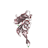
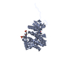
structures with the least restraint violations,structures with the lowest energy
Representative
Model #1
closest to the average
-
Components
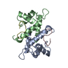
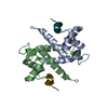
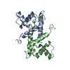
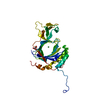
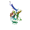
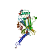
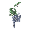
#1: Protein
S-100protein, betachain
Mass: 10550.776 Da / Num. of mol.: 2 Source method: isolated from a genetically manipulated source Source: (gene. exp.) Bos taurus (domestic cattle) / Gene: S100B / Plasmid: pH6 TEV / Production host: Escherichia coli (E. coli) / Strain (production host): BL21 Codon Plus (DE3) RIL / References: UniProt: P02638
#2: Protein/peptide
NdrSer/Thrkinase-likeprotein
Mass: 3232.855 Da / Num. of mol.: 2 Fragment: N-terminal regulatory domain fragment, sequence database residue 60-85 Source method: obtained synthetically / Details: This sequence occurs naturally in Homo sapiens. / References: UniProt: Q15208
-
Experimental details
-
Experiment
Experiment
Method: SOLUTION NMR
NMR experiment
Conditions-ID
Experiment-ID
Solution-ID
Type
1
1
1
3D 15N-separated NOESY
2
2
2
3D 13C-separated NOESY
3
3
3
3D 13C-separated NOESY
2
4
2
2D 15N/13C F2-Filtered NOESY
2
5
2
3D 13C-F1 separated 13C/15N-F2 Filtered HMQC-NOESY
NMR details
Text: Protein Backbone/Side-Chain Assignments were made from 3D CBCA(CO)NH, 3D HNCA, 3D HN(CO)CA, 3D HNCO 3D HCCH-TOCSY, 3D HCCH-COSY experiments. Peptide was assigned from 2D-Filtered COSY/TOCSY Experiments.
-
Sample preparation
Details
Solution-ID
Contents
Solvent system
1
1 mM 1H,15N labeled protein 1 mM unlabeled peptide 20 mM d-11 tris and 10mM d10-DTT 5 mM CaCl2 and 30mM KCL
90% H2O/10% D2O
2
1 mM 1H,13C,15N labeled protein 1 mM unlabeled peptide 20 mM d-11 tris and 10mM d10-DTT 5 mM CaCl2 and 30mM KCL
90% H2O/10% D2O
3
1 mM 1H,13C,15N labeled protein 1 mM unlabeled peptide 20 mM d-11 tris and 10mM d10-DTT 5 mM CaCl2 and 30mM KCL
100% D2O
Sample conditions
Conditions-ID
Ionic strength
pH
Pressure (kPa)
Temperature (K)
1
0.045M/L
7.5
ambient
310K
2
0.045M/L
7.5
ambient
310K
3
0.045M/L
7.5
ambient
310K
Crystal grow
*PLUS
Method: other / Details: NMR
-
NMR measurement
Radiation
Protocol: SINGLE WAVELENGTH / Monochromatic (M) / Laue (L): M
Method: Torsion Angle Dynamics, Restrained Molecular Dynamics, Simulated Annealing, Software ordinal: 1 Details: The structures are based on a total of 3274 restraints, 2964 are NOE-derived distance constraints and 310 dihedral angle restraints.
NMR representative
Selection criteria: closest to the average
NMR ensemble
Conformer selection criteria: structures with the least restraint violations,structures with the lowest energy Conformers calculated total number: 128 / Conformers submitted total number: 20
+
About Yorodumi
-
News
-
Feb 9, 2022. New format data for meta-information of EMDB entries
New format data for meta-information of EMDB entries
Version 3 of the EMDB header file is now the official format.
The previous official version 1.9 will be removed from the archive.
In the structure databanks used in Yorodumi, some data are registered as the other names, "COVID-19 virus" and "2019-nCoV". Here are the details of the virus and the list of structure data.
Jan 31, 2019. EMDB accession codes are about to change! (news from PDBe EMDB page)
EMDB accession codes are about to change! (news from PDBe EMDB page)
The allocation of 4 digits for EMDB accession codes will soon come to an end. Whilst these codes will remain in use, new EMDB accession codes will include an additional digit and will expand incrementally as the available range of codes is exhausted. The current 4-digit format prefixed with “EMD-” (i.e. EMD-XXXX) will advance to a 5-digit format (i.e. EMD-XXXXX), and so on. It is currently estimated that the 4-digit codes will be depleted around Spring 2019, at which point the 5-digit format will come into force.
The EM Navigator/Yorodumi systems omit the EMD- prefix.
Related info.:Q: What is EMD? / ID/Accession-code notation in Yorodumi/EM Navigator
Yorodumi is a browser for structure data from EMDB, PDB, SASBDB, etc.
This page is also the successor to EM Navigator detail page, and also detail information page/front-end page for Omokage search.
The word "yorodu" (or yorozu) is an old Japanese word meaning "ten thousand". "mi" (miru) is to see.
Related info.:EMDB / PDB / SASBDB / Comparison of 3 databanks / Yorodumi Search / Aug 31, 2016. New EM Navigator & Yorodumi / Yorodumi Papers / Jmol/JSmol / Function and homology information / Changes in new EM Navigator and Yorodumi
 Movie
Movie Controller

 Yorodumi
 Open data
 Basic information
 Components
 Keywords
 Function and homology information
 Authors
 Citation
 Structure visualization
 Downloads & links
 Other downloads
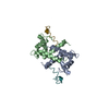


 PDBj
PDBj


 Assembly
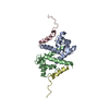

 Sample preparation
 Processing
 Amber
Amber