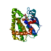
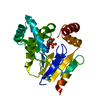
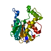
Method: HYBRID DISTANCE GEOMETRY, SIMULATED ANNEALING / Software ordinal: 1 Details: THE STRUCTURES OF THE MONOMERS WERE CALCULATED USING MATRIX DISTANCE GEOMETRY, FOLLOWED BY SIMULATED ANNEALING. THE MONOMERS WERE THEN DUPLICATED AND EACH NEW SUBUNIT WAS ROTATED 180 DEGREES ...Details: THE STRUCTURES OF THE MONOMERS WERE CALCULATED USING MATRIX DISTANCE GEOMETRY, FOLLOWED BY SIMULATED ANNEALING. THE MONOMERS WERE THEN DUPLICATED AND EACH NEW SUBUNIT WAS ROTATED 180 DEGREES AND SEPARATELY FROM ITS PARTNER BY 40 ANGSTROMS. NO DOCKING OF THE TWO MONOMERS WAS ATTEMPTED PRIOR THE FURTHER STRUCTURE CALCULATIONS. DIMER STRUCTURE CALCULATIONS USED THE 40 UNAMBIGUOUS INTERMONOMER NOES TO BRING THE SUBUNITS IN A POSITION RELATIVE TO ONE ANOTHER. A FURTHER FOUR ROUNDS OF SIMULATED ANNEALING GENERATED THE FINAL STRUCTURES. AT NO POINT DURING THE DIMER CALCULATIONS WAS NON-CRYSTALLOGRAPHIC SYMMETRY UTILIZED.
NMR ensemble
Conformer selection criteria: LEAST RESTRAINT VIOLATIONS / Conformers calculated total number: 30 / Conformers submitted total number: 20
+
About Yorodumi
-
News
-
Feb 9, 2022. New format data for meta-information of EMDB entries
New format data for meta-information of EMDB entries
Version 3 of the EMDB header file is now the official format.
The previous official version 1.9 will be removed from the archive.
In the structure databanks used in Yorodumi, some data are registered as the other names, "COVID-19 virus" and "2019-nCoV". Here are the details of the virus and the list of structure data.
Jan 31, 2019. EMDB accession codes are about to change! (news from PDBe EMDB page)
EMDB accession codes are about to change! (news from PDBe EMDB page)
The allocation of 4 digits for EMDB accession codes will soon come to an end. Whilst these codes will remain in use, new EMDB accession codes will include an additional digit and will expand incrementally as the available range of codes is exhausted. The current 4-digit format prefixed with “EMD-” (i.e. EMD-XXXX) will advance to a 5-digit format (i.e. EMD-XXXXX), and so on. It is currently estimated that the 4-digit codes will be depleted around Spring 2019, at which point the 5-digit format will come into force.
The EM Navigator/Yorodumi systems omit the EMD- prefix.
Related info.:Q: What is EMD? / ID/Accession-code notation in Yorodumi/EM Navigator
Yorodumi is a browser for structure data from EMDB, PDB, SASBDB, etc.
This page is also the successor to EM Navigator detail page, and also detail information page/front-end page for Omokage search.
The word "yorodu" (or yorozu) is an old Japanese word meaning "ten thousand". "mi" (miru) is to see.
Related info.:EMDB / PDB / SASBDB / Comparison of 3 databanks / Yorodumi Search / Aug 31, 2016. New EM Navigator & Yorodumi / Yorodumi Papers / Jmol/JSmol / Function and homology information / Changes in new EM Navigator and Yorodumi
 Movie
Movie Controller


 Open data
 Basic information
 Components
 Keywords
 Function and homology information
 Homo sapiens (human)
 Authors
 Citation
 Structure visualization
 Downloads & links
 Other downloads
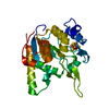
 PDBj
PDBj


 Assembly

 HSQC
HSQC Sample preparation
 Processing