 Movie
Movie Controller


+ Open data

- Basic information
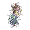
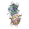
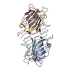
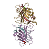
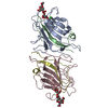
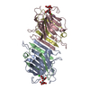
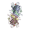
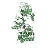
| Entry | Database: PDB / ID: 2qni | ||||||
|---|---|---|---|---|---|---|---|
| Title | Crystal structure of uncharacterized protein Atu0299 | ||||||
 Components Components | Uncharacterized protein Atu0299 | ||||||
 Keywords Keywords | STRUCTURAL GENOMICS / UNKNOWN FUNCTION / MCSG / Atu0299 / In situ proteolysis / PSI-2 / Protein Structure Initiative / Midwest Center for Structural Genomics / Uncharacterized protein | ||||||
| Function / homology | Phosphoglycerate mutase-like / Histidine phosphatase superfamily, clade-1 / Histidine phosphatase superfamily (branch 1) / Histidine phosphatase superfamily / Rossmann fold / 3-Layer(aba) Sandwich / Alpha Beta / Histidine phosphatase family protein / :  Function and homology information Function and homology information | ||||||
| Biological species |  Agrobacterium tumefaciens str. (bacteria) Agrobacterium tumefaciens str. (bacteria) | ||||||
| Method |  X-RAY DIFFRACTION / SYNCHROTRON / SAD / Resolution: 1.8 Å X-RAY DIFFRACTION / SYNCHROTRON / SAD / Resolution: 1.8 Å | ||||||
 Authors Authors | Dong, A. / Xu, X. / Gu, J. / Zheng, H. / Edwards, A.M. / Joachimiak, A. / Savchenko, A. / Midwest Center for Structural Genomics (MCSG) | ||||||
 Citation Citation | Journal: To be Published Title: Crystal structure of uncharacterized protein Atu0299. Authors: Dong, A. / Xu, X. / Gu, J. / Zheng, H. / Edwards, A.M. / Joachimiak, A. / Savchenko, A. | ||||||
| History |
| ||||||
| Remark 300 | BIOMOLECULE: 1 SEE REMARK 350 FOR THE AUTHOR PROVIDED AND PROGRAM GENERATED ASSEMBLY INFORMATION ... BIOMOLECULE: 1 SEE REMARK 350 FOR THE AUTHOR PROVIDED AND PROGRAM GENERATED ASSEMBLY INFORMATION FOR THE STRUCTURE IN THIS ENTRY. THE REMARK MAY ALSO PROVIDE INFORMATION ON BURIED SURFACE AREA. AUTHORS STATE THAT THE BIOLOGICAL UNIT INFORMATION OF THIS PROTEIN IS NOT AVAILABLE. |
- Structure visualization
| Structure viewer | Molecule: MolmilJmol/JSmol |
|---|
- Downloads & links
-Download
| PDBx/mmCIF format | 2qni.cif.gz | 55 KB | Display | PDBx/mmCIF format |
|---|---|---|---|---|
| PDB format | pdb2qni.ent.gz | 39.3 KB | Display | PDB format |
| PDBx/mmJSON format | 2qni.json.gz | Tree view | PDBx/mmJSON format | |
| Others |  Other downloads Other downloads |
-Validation report
| Arichive directory | https://data.pdbj.org/pub/pdb/validation_reports/qn/2qniftp://data.pdbj.org/pub/pdb/validation_reports/qn/2qni | HTTPS FTP |
|---|
-Related structure data
| Similar structure data | |
|---|---|
| Other databases |


-Links
 PDBj
PDBj


- Assembly
| Deposited unit | 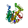
| ||||||||
|---|---|---|---|---|---|---|---|---|---|
| 1 | 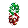
| ||||||||
| Unit cell |
|
-Components
| #1: Protein | Mass: 24388.555 Da / Num. of mol.: 1 Source method: isolated from a genetically manipulated source Source: (gene. exp.) Agrobacterium tumefaciens str. (bacteria)Species: Agrobacterium tumefaciens / Strain: C58 / Gene: AGR_C_517, Atu0299 / Plasmid: pET derivative / Production host: |
|---|---|
| #2: Water | ChemComp-HOH /  Mass: 18.015 Da / Num. of mol.: 177 / Source method: isolated from a natural source / Formula: H2O Mass: 18.015 Da / Num. of mol.: 177 / Source method: isolated from a natural source / Formula: H2O |
| Has protein modification | Y |
-Experimental details
-Experiment
| Experiment | Method: X-RAY DIFFRACTION / Number of used crystals: 1 |
|---|
- Sample preparation
| Crystal | Density Matthews: 3.33 Å3/Da / Density % sol: 63.03 % Description: Due to a number of circumstances, initial data processing was only accomplished in space group C2. Scaling statistics is provided for that space group. Using the program XPREP, the ...Description: Due to a number of circumstances, initial data processing was only accomplished in space group C2. Scaling statistics is provided for that space group. Using the program XPREP, the higher symmetry (H32) was detected and used in refinement. The structure factor file contains both data sets: for H32 and for C2 space group assignments |
|---|---|
| Crystal grow | Temperature: 297 K / Method: vapor diffusion, sitting drop / pH: 4.6 Details: 0.1M Na(OAC) pH 4.6, 3.5M Sodium formate. THE CRYSTAL WAS OBTAINED BY IN SITU PROTEOLYSIS, PROTEIN SAMPLE WAS MIXED WITH 1:100 WEIGHT TO WEIGHT RATIO WITH CHYMOTRYPSIN IMMEDIATELY PRIOR TO ...Details: 0.1M Na(OAC) pH 4.6, 3.5M Sodium formate. THE CRYSTAL WAS OBTAINED BY IN SITU PROTEOLYSIS, PROTEIN SAMPLE WAS MIXED WITH 1:100 WEIGHT TO WEIGHT RATIO WITH CHYMOTRYPSIN IMMEDIATELY PRIOR TO SETTING UP CRYSTALLIZATION, VAPOR DIFFUSION, SITTING DROP, temperature 297K Temp details: THE CRYSTAL WAS OBTAINED BY IN SITU PROTEOLYSIS, PROTEIN SAMPLE WAS MIXED WITH 1:100 WEIGHT TO WEIGHT RATIO WITH CHYMOTRYPSIN IMMEDIATELY PRIOR TO SETTING UP CRYSTALLIZATION |
-Data collection
| Diffraction | Mean temperature: 100 K |
|---|---|
| Diffraction source | Source: SYNCHROTRON / Site: CHESS  / Beamline: A1 / Wavelength: 0.976 Å / Beamline: A1 / Wavelength: 0.976 Å |
| Detector | Type: ADSC QUANTUM 210 / Detector: CCD / Date: May 5, 2007 |
| Radiation | Monochromator: Si(111) / Protocol: SINGLE WAVELENGTH / Monochromatic (M) / Laue (L): M / Scattering type: x-ray |
| Radiation wavelength | Wavelength: 0.976 Å / Relative weight: 1 |
| Reflection | Resolution: 1.8→50 Å / Num. all: 88206 / Num. obs: 88206 / % possible obs: 99.9 % / Observed criterion σ(F): 0 / Observed criterion σ(I): 0 / Redundancy: 7.5 % / Biso Wilson estimate: 28.4 Å2 / Rmerge(I) obs: 0.061 / Rsym value: 0.061 / Net I/σ(I): 10.9 |
| Reflection shell | Resolution: 1.8→1.86 Å / Redundancy: 6.1 % / Rmerge(I) obs: 0.749 / Mean I/σ(I) obs: 1.92 / Num. unique all: 8762 / Rsym value: 0.749 / % possible all: 99.3 |
- Processing
| Software |
| ||||||||||||||||||||||||||||||||||||||||||||||||||||||||||||||||||||||||||||||||||||||||||||||||||||||||||||||||||||||||||||||||||||||||||||||||||||||||||||||||||||||||||
|---|---|---|---|---|---|---|---|---|---|---|---|---|---|---|---|---|---|---|---|---|---|---|---|---|---|---|---|---|---|---|---|---|---|---|---|---|---|---|---|---|---|---|---|---|---|---|---|---|---|---|---|---|---|---|---|---|---|---|---|---|---|---|---|---|---|---|---|---|---|---|---|---|---|---|---|---|---|---|---|---|---|---|---|---|---|---|---|---|---|---|---|---|---|---|---|---|---|---|---|---|---|---|---|---|---|---|---|---|---|---|---|---|---|---|---|---|---|---|---|---|---|---|---|---|---|---|---|---|---|---|---|---|---|---|---|---|---|---|---|---|---|---|---|---|---|---|---|---|---|---|---|---|---|---|---|---|---|---|---|---|---|---|---|---|---|---|---|---|---|---|---|
| Refinement | Method to determine structure: SAD / Resolution: 1.8→50 Å / Cor.coef. Fo:Fc: 0.955 / Cor.coef. Fo:Fc free: 0.945 / SU B: 2.034 / SU ML: 0.065 / Cross valid method: THROUGHOUT / σ(F): 0 / σ(I): 0 / ESU R: 0.099 / ESU R Free: 0.096 / Stereochemistry target values: MAXIMUM LIKELIHOOD Details: HYDROGENS HAVE BEEN ADDED IN THE RIDING POSITIONS. ARP/wARP 6.1.1 and COOT 0.1 programs were also used in refinement
| ||||||||||||||||||||||||||||||||||||||||||||||||||||||||||||||||||||||||||||||||||||||||||||||||||||||||||||||||||||||||||||||||||||||||||||||||||||||||||||||||||||||||||
| Solvent computation | Ion probe radii: 0.8 Å / Shrinkage radii: 0.8 Å / VDW probe radii: 1.4 Å / Solvent model: MASK | ||||||||||||||||||||||||||||||||||||||||||||||||||||||||||||||||||||||||||||||||||||||||||||||||||||||||||||||||||||||||||||||||||||||||||||||||||||||||||||||||||||||||||
| Displacement parameters | Biso mean: 28.7 Å2
| ||||||||||||||||||||||||||||||||||||||||||||||||||||||||||||||||||||||||||||||||||||||||||||||||||||||||||||||||||||||||||||||||||||||||||||||||||||||||||||||||||||||||||
| Refinement step | Cycle: LAST / Resolution: 1.8→50 Å
| ||||||||||||||||||||||||||||||||||||||||||||||||||||||||||||||||||||||||||||||||||||||||||||||||||||||||||||||||||||||||||||||||||||||||||||||||||||||||||||||||||||||||||
| Refine LS restraints |
| ||||||||||||||||||||||||||||||||||||||||||||||||||||||||||||||||||||||||||||||||||||||||||||||||||||||||||||||||||||||||||||||||||||||||||||||||||||||||||||||||||||||||||
| LS refinement shell | Resolution: 1.8→1.85 Å / Total num. of bins used: 20
|
