 Movie
Movie Controller

[English] 日本語
 Yorodumi
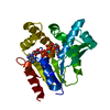
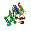
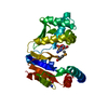
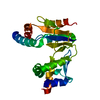
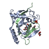
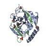
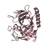
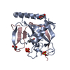
- PDB-7ckj: Crystal structure of CMP kinase in complex with CMP from Thermus ... -
+ Open data

- Basic information
| Entry | Database: PDB / ID: 7ckj | |||||||||
|---|---|---|---|---|---|---|---|---|---|---|
| Title | Crystal structure of CMP kinase in complex with CMP from Thermus thermophilus HB8 | |||||||||
 Components Components | Cytidylate kinase | |||||||||
 Keywords Keywords | TRANSFERASE / CMP kinase / CMP complex / open conformation / nucleotide metabolism / Structural Genomics / PSI-2 / Protein Structure Initiative / RIKEN Structural Genomics/Proteomics Initiative / RSGI | |||||||||
| Function / homology |  Function and homology information Function and homology information(d)CMP kinase / CMP kinase activity / dCMP kinase activity / pyrimidine nucleotide metabolic process / ATP binding / cytoplasm Similarity search - Function | |||||||||
| Biological species |   Thermus thermophilus HB8 (bacteria) Thermus thermophilus HB8 (bacteria) | |||||||||
| Method |  X-RAY DIFFRACTION / SYNCHROTRON / MOLECULAR REPLACEMENT / Resolution: 1.5 Å X-RAY DIFFRACTION / SYNCHROTRON / MOLECULAR REPLACEMENT / Resolution: 1.5 Å | |||||||||
 Authors Authors | Mega, R. / Nakagawa, N. / Kuramitsu, S. / Masui, R. / RIKEN Structural Genomics/Proteomics Initiative (RSGI) | |||||||||
| Funding support |  Japan, 2items Japan, 2items
| |||||||||
 Citation Citation | Journal: PLoS ONE / Year: 2020 Title: The crystal structures of Thermus thermophilus CMP kinase complexed with a phosphoryl group acceptor and donor. Authors: Mega, R. / Nakagawa, N. / Kuramitsu, S. / Masui, R. | |||||||||
| History |
|
- Structure visualization
| Structure viewer | Molecule: MolmilJmol/JSmol |
|---|
- Downloads & links
-Download
| PDBx/mmCIF format | 7ckj.cif.gz | 58.1 KB | Display | PDBx/mmCIF format |
|---|---|---|---|---|
| PDB format | pdb7ckj.ent.gz | 39.3 KB | Display | PDB format |
| PDBx/mmJSON format | 7ckj.json.gz | Tree view | PDBx/mmJSON format | |
| Others |  Other downloads Other downloads |
-Validation report
| Arichive directory | https://data.pdbj.org/pub/pdb/validation_reports/ck/7ckjftp://data.pdbj.org/pub/pdb/validation_reports/ck/7ckj | HTTPS FTP |
|---|
-Related structure data
| Related structure data | 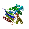 2cmkS S: Starting model for refinement |
|---|---|
| Similar structure data |


-Links
 PDBj
PDBj- Assembly
| Deposited unit | 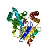
| ||||||||
|---|---|---|---|---|---|---|---|---|---|
| 1 |
| ||||||||
| Unit cell |
|
-Components
| #1: Protein | Mass: 22584.818 Da / Num. of mol.: 1 Source method: isolated from a genetically manipulated source Source: (gene. exp.) Thermus thermophilus HB8 (bacteria) / Strain: HB8 / Gene: cmk, TTHA0458 / Plasmid: pET-11a / Production host: |
|---|---|
| #2: Chemical | ChemComp-C5P /   Mass: 323.197 Da / Num. of mol.: 1 / Source method: obtained synthetically / Formula: C9H14N3O8P / Feature type: SUBJECT OF INVESTIGATION Mass: 323.197 Da / Num. of mol.: 1 / Source method: obtained synthetically / Formula: C9H14N3O8P / Feature type: SUBJECT OF INVESTIGATION |
| #3: Water | ChemComp-HOH /  Mass: 18.015 Da / Num. of mol.: 135 / Source method: isolated from a natural source / Formula: H2O Mass: 18.015 Da / Num. of mol.: 135 / Source method: isolated from a natural source / Formula: H2O |
| Has ligand of interest | Y |
-Experimental details
-Experiment
| Experiment | Method: X-RAY DIFFRACTION / Number of used crystals: 1 |
|---|
- Sample preparation
| Crystal | Density Matthews: 2.71 Å3/Da / Density % sol: 54.66 % |
|---|---|
| Crystal grow | Temperature: 293 K / Method: vapor diffusion, sitting drop / pH: 7 Details: 0.1M BIS-TRIS, 3.5M Sodium formate, pH 7.0, VAPOR DIFFUSION, SITTING DROP, temperature 293K |
-Data collection
| Diffraction | Mean temperature: 90 K / Serial crystal experiment: N | |||||||||||||||||||||||||||||||||||||||||||||||||||||||||||||||||||||||||||||
|---|---|---|---|---|---|---|---|---|---|---|---|---|---|---|---|---|---|---|---|---|---|---|---|---|---|---|---|---|---|---|---|---|---|---|---|---|---|---|---|---|---|---|---|---|---|---|---|---|---|---|---|---|---|---|---|---|---|---|---|---|---|---|---|---|---|---|---|---|---|---|---|---|---|---|---|---|---|---|
| Diffraction source | Source: SYNCHROTRON / Site: SPring-8 / Beamline: BL45XU / Wavelength: 1 Å | |||||||||||||||||||||||||||||||||||||||||||||||||||||||||||||||||||||||||||||
| Detector | Type: ADSC QUANTUM 210 / Detector: CCD / Date: Jul 19, 2006 | |||||||||||||||||||||||||||||||||||||||||||||||||||||||||||||||||||||||||||||
| Radiation | Monochromator: transparent diamond double crystal / Protocol: SINGLE WAVELENGTH / Monochromatic (M) / Laue (L): M / Scattering type: x-ray | |||||||||||||||||||||||||||||||||||||||||||||||||||||||||||||||||||||||||||||
| Radiation wavelength | Wavelength: 1 Å / Relative weight: 1 | |||||||||||||||||||||||||||||||||||||||||||||||||||||||||||||||||||||||||||||
| Reflection | Resolution: 1.5→50 Å / Num. obs: 38410 / % possible obs: 99.9 % / Redundancy: 8.1 % / Rmerge(I) obs: 0.066 / Χ2: 1.997 / Net I/σ(I): 28 / Num. measured all: 312320 | |||||||||||||||||||||||||||||||||||||||||||||||||||||||||||||||||||||||||||||
| Reflection shell |
|
- Processing
| Software |
| ||||||||||||||||||||||||
|---|---|---|---|---|---|---|---|---|---|---|---|---|---|---|---|---|---|---|---|---|---|---|---|---|---|
| Refinement | Method to determine structure: MOLECULAR REPLACEMENT Starting model: 2CMK Resolution: 1.5→50 Å / Cross valid method: THROUGHOUT / σ(F): 0
| ||||||||||||||||||||||||
| Solvent computation | Bsol: 45.9207 Å2 | ||||||||||||||||||||||||
| Displacement parameters | Biso max: 53.42 Å2 / Biso mean: 19.4958 Å2 / Biso min: 8.36 Å2
| ||||||||||||||||||||||||
| Refinement step | Cycle: final / Resolution: 1.5→50 Å
| ||||||||||||||||||||||||
| Refine LS restraints |
| ||||||||||||||||||||||||
| LS refinement shell | Resolution: 1.5→1.59 Å / Rfactor Rfree error: 0.001
| ||||||||||||||||||||||||
| Xplor file |
|
